 Movie
Movie Controller

[English] 日本語
 Yorodumi
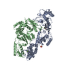
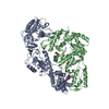
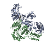
- PDB-7so4: Crystal Structure of HIV-1 Y181C mutant Reverse Transcriptase in ... -
+ Open data

- Basic information
| Entry | Database: PDB / ID: 7so4 | ||||||||||||
|---|---|---|---|---|---|---|---|---|---|---|---|---|---|
| Title | Crystal Structure of HIV-1 Y181C mutant Reverse Transcriptase in Complex with 5-(2-(2-(2,4-dioxo-3,4-dihydropyrimidin-1(2H)-yl)ethoxy)phenoxy)-7-fluoro-2-naphthonitrile (JLJ635), a Non-nucleoside Inhibitor | ||||||||||||
 Components Components |
| ||||||||||||
 Keywords Keywords | hydrolase / transferase/viral protein / NNRTIs / HIV-1 / drug design / TRANSFERASE / transferase-viral protein complex | ||||||||||||
| Function / homology |  Function and homology information Function and homology informationHIV-1 retropepsin / symbiont-mediated activation of host apoptosis / retroviral ribonuclease H / exoribonuclease H / exoribonuclease H activity / DNA integration / viral genome integration into host DNA / establishment of integrated proviral latency / RNA-directed DNA polymerase / RNA stem-loop binding ...HIV-1 retropepsin / symbiont-mediated activation of host apoptosis / retroviral ribonuclease H / exoribonuclease H / exoribonuclease H activity / DNA integration / viral genome integration into host DNA / establishment of integrated proviral latency / RNA-directed DNA polymerase / RNA stem-loop binding / viral penetration into host nucleus / host multivesicular body / RNA-directed DNA polymerase activity / RNA-DNA hybrid ribonuclease activity / Transferases; Transferring phosphorus-containing groups; Nucleotidyltransferases / host cell / viral nucleocapsid / DNA recombination / DNA-directed DNA polymerase / aspartic-type endopeptidase activity / Hydrolases; Acting on ester bonds / DNA-directed DNA polymerase activity / symbiont-mediated suppression of host gene expression / viral translational frameshifting / symbiont entry into host cell / lipid binding / host cell nucleus / host cell plasma membrane / virion membrane / structural molecule activity / proteolysis / DNA binding / zinc ion binding Similarity search - Function | ||||||||||||
| Biological species |  Human immunodeficiency virus type 1 group M subtype B Human immunodeficiency virus type 1 group M subtype B | ||||||||||||
| Method |  X-RAY DIFFRACTION / SYNCHROTRON / MOLECULAR REPLACEMENT / Resolution: 2.95 Å X-RAY DIFFRACTION / SYNCHROTRON / MOLECULAR REPLACEMENT / Resolution: 2.95 Å | ||||||||||||
 Authors Authors | Bertoletti, N. / Anderson, K.S. / Cisneros Trigo, J.A. / Jorgensen, W.L. / Frey, K.M. / Chan, A.H. | ||||||||||||
| Funding support |  United States, 3items United States, 3items
| ||||||||||||
 Citation Citation | Journal: Front Mol Biosci / Year: 2022 Title: Structural Studies and Structure Activity Relationships for Novel Computationally Designed Non-nucleoside Inhibitors and Their Interactions With HIV-1 Reverse Transcriptase. Authors: Frey, K.M. / Bertoletti, N. / Chan, A.H. / Ippolito, J.A. / Bollini, M. / Spasov, K.A. / Jorgensen, W.L. / Anderson, K.S. | ||||||||||||
| History |
|
- Structure visualization
| Structure viewer | Molecule: MolmilJmol/JSmol |
|---|
- Downloads & links
-Download
| PDBx/mmCIF format | 7so4.cif.gz | 207.4 KB | Display | PDBx/mmCIF format |
|---|---|---|---|---|
| PDB format | pdb7so4.ent.gz | 160.3 KB | Display | PDB format |
| PDBx/mmJSON format | 7so4.json.gz | Tree view | PDBx/mmJSON format | |
| Others |  Other downloads Other downloads |
-Validation report
| Arichive directory | https://data.pdbj.org/pub/pdb/validation_reports/so/7so4ftp://data.pdbj.org/pub/pdb/validation_reports/so/7so4 | HTTPS FTP |
|---|
-Related structure data
| Related structure data | 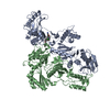 7snpC 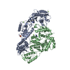 7snzC 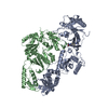 7so1C 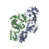 7so2C 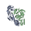 7so3C  7so6C  6oe3S S: Starting model for refinement C: citing same article ( |
|---|---|
| Similar structure data |


-Links
 PDBj
PDBj


- Assembly
| Deposited unit | 
| ||||||||||||
|---|---|---|---|---|---|---|---|---|---|---|---|---|---|
| 1 |
| ||||||||||||
| Unit cell |
|
-Components
| #1: Protein | Mass: 63929.207 Da / Num. of mol.: 1 / Mutation: Y181C Source method: isolated from a genetically manipulated source Source: (gene. exp.) Human immunodeficiency virus type 1 group M subtype BGene: gag-pol Production host:  References: UniProt: P03366, RNA-directed DNA polymerase, DNA-directed DNA polymerase, retroviral ribonuclease H, exoribonuclease H |
|---|---|
| #2: Protein | Mass: 50039.488 Da / Num. of mol.: 1 Source method: isolated from a genetically manipulated source Source: (gene. exp.) Human immunodeficiency virus type 1 group M subtype B (isolate BH10)Strain: isolate BH10 / Gene: gag-pol Production host: References: UniProt: P03366 |
| #3: Chemical | ChemComp-M9A /   Mass: 417.389 Da / Num. of mol.: 1 / Source method: obtained synthetically / Formula: C23H16FN3O4 / Feature type: SUBJECT OF INVESTIGATION Mass: 417.389 Da / Num. of mol.: 1 / Source method: obtained synthetically / Formula: C23H16FN3O4 / Feature type: SUBJECT OF INVESTIGATION |
| #4: Chemical | ChemComp-SO4 /   Mass: 96.063 Da / Num. of mol.: 1 / Source method: obtained synthetically / Formula: SO4 Mass: 96.063 Da / Num. of mol.: 1 / Source method: obtained synthetically / Formula: SO4 |
| #5: Water | ChemComp-HOH /  Mass: 18.015 Da / Num. of mol.: 3 / Source method: isolated from a natural source / Formula: H2O Mass: 18.015 Da / Num. of mol.: 3 / Source method: isolated from a natural source / Formula: H2O |
| Has ligand of interest | Y |
-Experimental details
-Experiment
| Experiment | Method: X-RAY DIFFRACTION / Number of used crystals: 1 |
|---|
- Sample preparation
| Crystal | Density Matthews: 2.75 Å3/Da / Density % sol: 55.33 % |
|---|---|
| Crystal grow | Temperature: 277 K / Method: vapor diffusion, hanging drop / pH: 6 Details: 50 mM MES pH 6.0, 18% PEG 8,000, 100 mM ammonium sulfate, 15 mM magnesium sulfate, and 5 mM spermine |
-Data collection
| Diffraction | Mean temperature: 100 K / Serial crystal experiment: N |
|---|---|
| Diffraction source | Source: SYNCHROTRON / Site: APS / Beamline: 24-ID-E / Wavelength: 0.97918 Å |
| Detector | Type: DECTRIS EIGER X 16M / Detector: PIXEL / Date: Jun 24, 2015 |
| Radiation | Protocol: SINGLE WAVELENGTH / Monochromatic (M) / Laue (L): M / Scattering type: x-ray |
| Radiation wavelength | Wavelength: 0.97918 Å / Relative weight: 1 |
| Reflection | Resolution: 2.95→50 Å / Num. obs: 26134 / % possible obs: 98.7 % / Redundancy: 3.7 % / Biso Wilson estimate: 82.38 Å2 / CC1/2: 0.999 / Rrim(I) all: 0.069 / Rsym value: 0.059 / Net I/σ(I): 17.82 |
| Reflection shell | Resolution: 2.95→3.12 Å / Redundancy: 4 % / Mean I/σ(I) obs: 2.41 / Num. unique obs: 3988 / CC1/2: 0.807 / CC star: 0.807 / Rrim(I) all: 0.74 / Rsym value: 0.63 / % possible all: 94.7 |
- Processing
| Software |
| ||||||||||||||||||||||||||||||||||||||||||||||||||||||||||||||||||||||
|---|---|---|---|---|---|---|---|---|---|---|---|---|---|---|---|---|---|---|---|---|---|---|---|---|---|---|---|---|---|---|---|---|---|---|---|---|---|---|---|---|---|---|---|---|---|---|---|---|---|---|---|---|---|---|---|---|---|---|---|---|---|---|---|---|---|---|---|---|---|---|---|
| Refinement | Method to determine structure: MOLECULAR REPLACEMENT Starting model: 6OE3 Resolution: 2.95→43.02 Å / SU ML: 0.4479 / Cross valid method: FREE R-VALUE / σ(F): 0.83 / Phase error: 30.9357 Stereochemistry target values: GeoStd + Monomer Library + CDL v1.2
| ||||||||||||||||||||||||||||||||||||||||||||||||||||||||||||||||||||||
| Solvent computation | Shrinkage radii: 0.9 Å / VDW probe radii: 1.11 Å / Solvent model: FLAT BULK SOLVENT MODEL | ||||||||||||||||||||||||||||||||||||||||||||||||||||||||||||||||||||||
| Displacement parameters | Biso mean: 89.02 Å2 | ||||||||||||||||||||||||||||||||||||||||||||||||||||||||||||||||||||||
| Refinement step | Cycle: LAST / Resolution: 2.95→43.02 Å
| ||||||||||||||||||||||||||||||||||||||||||||||||||||||||||||||||||||||
| Refine LS restraints |
| ||||||||||||||||||||||||||||||||||||||||||||||||||||||||||||||||||||||
| LS refinement shell |
|
