 Movie
Movie Controller

[English] 日本語
 Yorodumi
- PDB-7skj: Crystal structure of chloroplast triosephosphate isomerase from C... -
+ Open data

- Basic information
| Entry | Database: PDB / ID: 7skj | ||||||
|---|---|---|---|---|---|---|---|
| Title | Crystal structure of chloroplast triosephosphate isomerase from Cuscuta australis | ||||||
 Components Components | Triosephosphate isomerase | ||||||
 Keywords Keywords | ISOMERASE / TIM / triosephosphate isomerase / glycolysis / gluconeogenesis / Calvin-Benson-Bassham Cycle | ||||||
| Function / homology |  Function and homology information Function and homology informationtriose-phosphate isomerase activity / glyceraldehyde-3-phosphate biosynthetic process / glycerol catabolic process / glycolytic process / gluconeogenesis / cytosol Similarity search - Function | ||||||
| Biological species |  Cuscuta australis (plant) Cuscuta australis (plant) | ||||||
| Method |  X-RAY DIFFRACTION / SYNCHROTRON / MOLECULAR REPLACEMENT / Resolution: 1.9 Å X-RAY DIFFRACTION / SYNCHROTRON / MOLECULAR REPLACEMENT / Resolution: 1.9 Å | ||||||
 Authors Authors | Jones, G. / Vickers, C. / Patrick, W. | ||||||
| Funding support |  New Zealand, 1items New Zealand, 1items
| ||||||
 Citation Citation | Journal: To Be Published Title: Crystal structure of chloroplast triosephosphate isomerase from Cuscuta australis Authors: Jones, G. / Vickers, C. / Patrick, W. | ||||||
| History |
|
- Structure visualization
| Structure viewer | Molecule: MolmilJmol/JSmol |
|---|
- Downloads & links
-Download
| PDBx/mmCIF format | 7skj.cif.gz | 256.7 KB | Display | PDBx/mmCIF format |
|---|---|---|---|---|
| PDB format | pdb7skj.ent.gz | 162.9 KB | Display | PDB format |
| PDBx/mmJSON format | 7skj.json.gz | Tree view | PDBx/mmJSON format | |
| Others |  Other downloads Other downloads |
-Validation report
| Arichive directory | https://data.pdbj.org/pub/pdb/validation_reports/sk/7skjftp://data.pdbj.org/pub/pdb/validation_reports/sk/7skj | HTTPS FTP |
|---|
-Related structure data
| Related structure data | 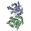 4ohqS S: Starting model for refinement |
|---|---|
| Similar structure data |
-Links
 PDBj
PDBj
- Assembly
| Deposited unit | 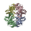
| ||||||||||||
|---|---|---|---|---|---|---|---|---|---|---|---|---|---|
| 1 | 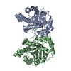
| ||||||||||||
| 2 | 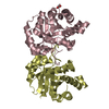
| ||||||||||||
| Unit cell |
|
-Components
| #1: Protein | Mass: 29568.379 Da / Num. of mol.: 4 Source method: isolated from a genetically manipulated source Source: (gene. exp.) Cuscuta australis (plant) / Gene: DM860_005218 / Production host:  #2: Chemical |   Mass: 59.044 Da / Num. of mol.: 2 / Source method: obtained synthetically / Formula: C2H3O2 / Feature type: SUBJECT OF INVESTIGATION Mass: 59.044 Da / Num. of mol.: 2 / Source method: obtained synthetically / Formula: C2H3O2 / Feature type: SUBJECT OF INVESTIGATION#3: Water | ChemComp-HOH / |  Mass: 18.015 Da / Num. of mol.: 892 / Source method: isolated from a natural source / Formula: H2O Mass: 18.015 Da / Num. of mol.: 892 / Source method: isolated from a natural source / Formula: H2OHas ligand of interest | Y | |
|---|
-Experimental details
-Experiment
| Experiment | Method: X-RAY DIFFRACTION / Number of used crystals: 1 |
|---|
- Sample preparation
| Crystal | Density Matthews: 2.01 Å3/Da / Density % sol: 38.7 % |
|---|---|
| Crystal grow | Temperature: 291.15 K / Method: vapor diffusion, sitting drop Details: 0.2 M Ammonium acetate, 0.1 M HEPES pH 7.5, 25% w/v PEG 3350 |
-Data collection
| Diffraction | Mean temperature: 100 K / Serial crystal experiment: N |
|---|---|
| Diffraction source | Source: SYNCHROTRON / Site: Australian Synchrotron  / Beamline: MX2 / Wavelength: 0.9464 Å / Beamline: MX2 / Wavelength: 0.9464 Å |
| Detector | Type: ADSC QUANTUM 315r / Detector: CCD / Date: Aug 5, 2021 |
| Radiation | Protocol: SINGLE WAVELENGTH / Monochromatic (M) / Laue (L): M / Scattering type: x-ray |
| Radiation wavelength | Wavelength: 0.9464 Å / Relative weight: 1 |
| Reflection | Resolution: 1.9→44.97 Å / Num. obs: 75939 / % possible obs: 99.8 % / Redundancy: 13.6 % / Biso Wilson estimate: 24.25 Å2 / CC1/2: 0.999 / Net I/σ(I): 16.3 |
| Reflection shell | Resolution: 1.9→1.94 Å / Num. unique obs: 4271 / CC1/2: 0.928 |
- Processing
| Software |
| |||||||||||||||||||||||||||||||||||||||||||||||||||||||||||||||||||||||||||||||||||||||||||||||||||||||||
|---|---|---|---|---|---|---|---|---|---|---|---|---|---|---|---|---|---|---|---|---|---|---|---|---|---|---|---|---|---|---|---|---|---|---|---|---|---|---|---|---|---|---|---|---|---|---|---|---|---|---|---|---|---|---|---|---|---|---|---|---|---|---|---|---|---|---|---|---|---|---|---|---|---|---|---|---|---|---|---|---|---|---|---|---|---|---|---|---|---|---|---|---|---|---|---|---|---|---|---|---|---|---|---|---|---|---|
| Refinement | Method to determine structure: MOLECULAR REPLACEMENT Starting model: 4OHQ Resolution: 1.9→44.97 Å / SU ML: 0.2141 / Cross valid method: FREE R-VALUE / Phase error: 22.4965 Stereochemistry target values: GeoStd + Monomer Library + CDL v1.2
| |||||||||||||||||||||||||||||||||||||||||||||||||||||||||||||||||||||||||||||||||||||||||||||||||||||||||
| Solvent computation | Shrinkage radii: 0.9 Å / VDW probe radii: 1.11 Å / Solvent model: FLAT BULK SOLVENT MODEL | |||||||||||||||||||||||||||||||||||||||||||||||||||||||||||||||||||||||||||||||||||||||||||||||||||||||||
| Displacement parameters | Biso mean: 27.53 Å2 | |||||||||||||||||||||||||||||||||||||||||||||||||||||||||||||||||||||||||||||||||||||||||||||||||||||||||
| Refinement step | Cycle: LAST / Resolution: 1.9→44.97 Å
| |||||||||||||||||||||||||||||||||||||||||||||||||||||||||||||||||||||||||||||||||||||||||||||||||||||||||
| Refine LS restraints |
| |||||||||||||||||||||||||||||||||||||||||||||||||||||||||||||||||||||||||||||||||||||||||||||||||||||||||
| LS refinement shell |
|
