 Movie
Movie Controller


+ Open data

- Basic information
| Entry | Database: PDB / ID: 7sj5 | |||||||||
|---|---|---|---|---|---|---|---|---|---|---|
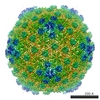
| Title | Bacteriophage lambda major capsid protein mutant - W308A | |||||||||
 Components Components | Major capsid protein | |||||||||
 Keywords Keywords | STRUCTURAL PROTEIN / major capsid protein / HK97-fold / assembly incompetent | |||||||||
| Function / homology |  Function and homology information Function and homology information | |||||||||
| Biological species |  Escherichia phage lambda (virus) Escherichia phage lambda (virus) | |||||||||
| Method |  X-RAY DIFFRACTION / SYNCHROTRON / MOLECULAR REPLACEMENT / Resolution: 2.695 Å X-RAY DIFFRACTION / SYNCHROTRON / MOLECULAR REPLACEMENT / Resolution: 2.695 Å | |||||||||
 Authors Authors | Davis, C.R. / Churchill, M.E. | |||||||||
| Funding support |  United States, 2items United States, 2items
| |||||||||
 Citation Citation | Journal: J.Mol.Biol. / Year: 2022 Title: Characterization of a Primordial Major Capsid-Scaffolding Protein Complex in Icosahedral Virus Shell Assembly. Authors: Davis, C.R. / Backos, D. / Morais, M.C. / Churchill, M.E.A. / Catalano, C.E. | |||||||||
| History |
|
- Structure visualization
| Structure viewer | Molecule: MolmilJmol/JSmol |
|---|
- Downloads & links
-Download
| PDBx/mmCIF format | 7sj5.cif.gz | 569 KB | Display | PDBx/mmCIF format |
|---|---|---|---|---|
| PDB format | pdb7sj5.ent.gz | 452.9 KB | Display | PDB format |
| PDBx/mmJSON format | 7sj5.json.gz | Tree view | PDBx/mmJSON format | |
| Others |  Other downloads Other downloads |
-Validation report
| Arichive directory | https://data.pdbj.org/pub/pdb/validation_reports/sj/7sj5ftp://data.pdbj.org/pub/pdb/validation_reports/sj/7sj5 | HTTPS FTP |
|---|
-Related structure data
| Related structure data |  3bqwS S: Starting model for refinement |
|---|---|
| Similar structure data |

-Links
 PDBj
PDBj
- Assembly
| Deposited unit | 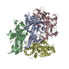
| ||||||||||||
|---|---|---|---|---|---|---|---|---|---|---|---|---|---|
| 1 | 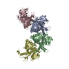
| ||||||||||||
| Unit cell |
|
-Components
| #1: Protein | Mass: 38114.027 Da / Num. of mol.: 4 / Mutation: W308A Source method: isolated from a genetically manipulated source Source: (gene. exp.) Escherichia phage lambda (virus) / Gene: E, lambdap08, gpE / Production host:  #2: Water | ChemComp-HOH / |  Mass: 18.015 Da / Num. of mol.: 163 / Source method: isolated from a natural source / Formula: H2O Mass: 18.015 Da / Num. of mol.: 163 / Source method: isolated from a natural source / Formula: H2O |
|---|
-Experimental details
-Experiment
| Experiment | Method: X-RAY DIFFRACTION / Number of used crystals: 1 |
|---|
- Sample preparation
| Crystal | Density Matthews: 2.37 Å3/Da / Density % sol: 48.17 % / Description: 70 micron rhomboid prism |
|---|---|
| Crystal grow | Temperature: 291.15 K / Method: vapor diffusion, hanging drop / pH: 5.7 Details: 0.1 M bis-tris, 0.1 M ammonium sulfate, 5% v/v glycerol, 30% v/v PEG-3350 |
-Data collection
| Diffraction | Mean temperature: 100 K / Serial crystal experiment: N |
|---|---|
| Diffraction source | Source: SYNCHROTRON / Site: ALS / Beamline: 4.2.2 / Wavelength: 1 Å |
| Detector | Type: CUSTOM-MADE / Detector: CCD / Date: Sep 30, 2017 |
| Radiation | Protocol: SINGLE WAVELENGTH / Monochromatic (M) / Laue (L): M / Scattering type: x-ray |
| Radiation wavelength | Wavelength: 1 Å / Relative weight: 1 |
| Reflection | Resolution: 2.695→29.75 Å / Num. obs: 39376 / % possible obs: 99.53 % / Redundancy: 3.7 % / Biso Wilson estimate: 49.19 Å2 / CC1/2: 0.999 / CC star: 1 / Rmerge(I) obs: 0.05594 / Rpim(I) all: 0.0336 / Rrim(I) all: 0.06535 / Net I/av σ(I): 18.27 / Net I/σ(I): 18.27 |
| Reflection shell | Resolution: 2.695→2.791 Å / Num. unique obs: 3819 / CC1/2: 0.919 |
- Processing
| Software |
| ||||||||||||||||||||||||
|---|---|---|---|---|---|---|---|---|---|---|---|---|---|---|---|---|---|---|---|---|---|---|---|---|---|
| Refinement | Method to determine structure: MOLECULAR REPLACEMENT Starting model: 3BQW Resolution: 2.695→29.75 Å / Cross valid method: FREE R-VALUE Stereochemistry target values: GeoStd + Monomer Library + CDL v1.2
| ||||||||||||||||||||||||
| Displacement parameters | Biso mean: 74.44 Å2 | ||||||||||||||||||||||||
| Refinement step | Cycle: LAST / Resolution: 2.695→29.75 Å
| ||||||||||||||||||||||||
| Refine LS restraints |
| ||||||||||||||||||||||||
| LS refinement shell | Resolution: 2.695→2.791 Å
|
