 Movie
Movie Controller

[English] 日本語
 Yorodumi
- PDB-7shq: Structure of a functional construct of eukaryotic elongation fact... -
+ Open data

- Basic information
| Entry | Database: PDB / ID: 7shq | ||||||
|---|---|---|---|---|---|---|---|
| Title | Structure of a functional construct of eukaryotic elongation factor 2 kinase in complex with calmodulin. | ||||||
 Components Components |
| ||||||
 Keywords Keywords | TRANSLATION / elongation factor 2 kinase / eEF2 / Calmodulin | ||||||
| Function / homology |  Function and homology information Function and homology informationelongation factor 2 kinase / elongation factor-2 kinase activity / regulation of translation at postsynapse / response to prolactin / myosin II filament disassembly / cellular response to anoxia / translation factor activity, RNA binding / positive regulation of dendritic spine morphogenesis / positive regulation of synapse assembly / CaM pathway ...elongation factor 2 kinase / elongation factor-2 kinase activity / regulation of translation at postsynapse / response to prolactin / myosin II filament disassembly / cellular response to anoxia / translation factor activity, RNA binding / positive regulation of dendritic spine morphogenesis / positive regulation of synapse assembly / CaM pathway / Cam-PDE 1 activation / Sodium/Calcium exchangers / Calmodulin induced events / Reduction of cytosolic Ca++ levels / Activation of Ca-permeable Kainate Receptor / CREB1 phosphorylation through the activation of CaMKII/CaMKK/CaMKIV cascasde / Loss of phosphorylation of MECP2 at T308 / CREB1 phosphorylation through the activation of Adenylate Cyclase / negative regulation of high voltage-gated calcium channel activity / PKA activation / CaMK IV-mediated phosphorylation of CREB / Glycogen breakdown (glycogenolysis) / CLEC7A (Dectin-1) induces NFAT activation / Activation of RAC1 downstream of NMDARs / negative regulation of ryanodine-sensitive calcium-release channel activity / organelle localization by membrane tethering / mitochondrion-endoplasmic reticulum membrane tethering / autophagosome membrane docking / negative regulation of calcium ion export across plasma membrane / regulation of cardiac muscle cell action potential / presynaptic endocytosis / Synthesis of IP3 and IP4 in the cytosol / regulation of cell communication by electrical coupling involved in cardiac conduction / Phase 0 - rapid depolarisation / calcineurin-mediated signaling / Negative regulation of NMDA receptor-mediated neuronal transmission / Unblocking of NMDA receptors, glutamate binding and activation / RHO GTPases activate PAKs / regulation of ryanodine-sensitive calcium-release channel activity / Ion transport by P-type ATPases / Uptake and function of anthrax toxins / mTORC1-mediated signalling / Long-term potentiation / protein phosphatase activator activity / Calcineurin activates NFAT / Regulation of MECP2 expression and activity / positive regulation of endocytosis / translational elongation / DARPP-32 events / Smooth Muscle Contraction / detection of calcium ion / regulation of cardiac muscle contraction / catalytic complex / RHO GTPases activate IQGAPs / regulation of cardiac muscle contraction by regulation of the release of sequestered calcium ion / calcium channel inhibitor activity / Activation of AMPK downstream of NMDARs / presynaptic cytosol / cellular response to interferon-beta / Protein methylation / regulation of release of sequestered calcium ion into cytosol by sarcoplasmic reticulum / cellular response to brain-derived neurotrophic factor stimulus / titin binding / Ion homeostasis / eNOS activation / Tetrahydrobiopterin (BH4) synthesis, recycling, salvage and regulation / regulation of calcium-mediated signaling / voltage-gated potassium channel complex / FCERI mediated Ca+2 mobilization / calcium channel complex / substantia nigra development / regulation of heart rate / Ras activation upon Ca2+ influx through NMDA receptor / FCGR3A-mediated IL10 synthesis / cellular response to calcium ion / Antigen activates B Cell Receptor (BCR) leading to generation of second messengers / cellular response to cAMP / calyx of Held / adenylate cyclase activator activity / sarcomere / VEGFR2 mediated cell proliferation / response to ischemia / protein serine/threonine kinase activator activity / VEGFR2 mediated vascular permeability / regulation of cytokinesis / spindle microtubule / positive regulation of receptor signaling pathway via JAK-STAT / Translocation of SLC2A4 (GLUT4) to the plasma membrane / calcium channel regulator activity / RAF activation / Transcriptional activation of mitochondrial biogenesis / response to calcium ion / cellular response to type II interferon / G2/M transition of mitotic cell cycle / Stimuli-sensing channels / cellular response to insulin stimulus / spindle pole / calcium-dependent protein binding / Signaling by RAF1 mutants / RAS processing Similarity search - Function | ||||||
| Biological species |  Homo sapiens (human) Homo sapiens (human) | ||||||
| Method |  X-RAY DIFFRACTION / SYNCHROTRON / MOLECULAR REPLACEMENT / Resolution: 2.34 Å X-RAY DIFFRACTION / SYNCHROTRON / MOLECULAR REPLACEMENT / Resolution: 2.34 Å | ||||||
 Authors Authors | Piserchio, A. / Isiorho, E.A. / Jeruzalmi, D. / Dalby, K.N. / Ghose, R. | ||||||
| Funding support |  United States, 1items United States, 1items
| ||||||
 Citation Citation | Journal: Sci Adv / Year: 2022 Title: Structural basis for the calmodulin-mediated activation of eukaryotic elongation factor 2 kinase. Authors: Piserchio, A. / Isiorho, E.A. / Long, K. / Bohanon, A.L. / Kumar, E.A. / Will, N. / Jeruzalmi, D. / Dalby, K.N. / Ghose, R. #1: Journal: Biorxiv / Year: 2022Title: Structural Basis for the Calmodulin-Mediated Activation of eEF-2K Authors: Piserchio, A. / Isiorho, E.A. / Long, K. / Bohanon, A.L. / Kumar, E.A. / Will, N. / Jeruzalmi, D. / Dalby, K.N. / Ghose, R. | ||||||
| History |
|
- Structure visualization
| Structure viewer | Molecule: MolmilJmol/JSmol |
|---|
- Downloads & links
-Download
| PDBx/mmCIF format | 7shq.cif.gz | 172.3 KB | Display | PDBx/mmCIF format |
|---|---|---|---|---|
| PDB format | pdb7shq.ent.gz | 113 KB | Display | PDB format |
| PDBx/mmJSON format | 7shq.json.gz | Tree view | PDBx/mmJSON format | |
| Others |  Other downloads Other downloads |
-Validation report
| Arichive directory | https://data.pdbj.org/pub/pdb/validation_reports/sh/7shqftp://data.pdbj.org/pub/pdb/validation_reports/sh/7shq | HTTPS FTP |
|---|
-Related structure data
| Related structure data |  1ia9S  3lkmS  3pdtS  3rjvS 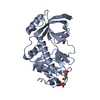 4kujS  4ozsS 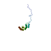 6nx4S S: Starting model for refinement |
|---|---|
| Similar structure data |
-Links
 PDBj
PDBj


- Assembly
| Deposited unit | 
| ||||||||||||
|---|---|---|---|---|---|---|---|---|---|---|---|---|---|
| 1 |
| ||||||||||||
| Unit cell |
| ||||||||||||
| Components on special symmetry positions |
|
-Components
-Protein , 2 types, 2 molecules AB
| #1: Protein | Mass: 60380.727 Da / Num. of mol.: 1 Source method: isolated from a genetically manipulated source Source: (gene. exp.) Homo sapiens (human) / Gene: EEF2K / Production host:  |
|---|---|
| #2: Protein | Mass: 16721.350 Da / Num. of mol.: 1 Source method: isolated from a genetically manipulated source Source: (gene. exp.) Homo sapiens (human) / Gene: CALM1, CALM, CAM, CAM1 / Production host: |
-Non-polymers , 4 types, 151 molecules 


| #3: Chemical | ChemComp-ZN /  Mass: 65.409 Da / Num. of mol.: 1 / Source method: obtained synthetically / Formula: Zn / Feature type: SUBJECT OF INVESTIGATION Mass: 65.409 Da / Num. of mol.: 1 / Source method: obtained synthetically / Formula: Zn / Feature type: SUBJECT OF INVESTIGATION | ||||
|---|---|---|---|---|---|
| #4: Chemical | ChemComp-MG /  Mass: 24.305 Da / Num. of mol.: 5 / Source method: obtained synthetically / Formula: Mg / Feature type: SUBJECT OF INVESTIGATION Mass: 24.305 Da / Num. of mol.: 5 / Source method: obtained synthetically / Formula: Mg / Feature type: SUBJECT OF INVESTIGATION#5: Chemical |  Mass: 40.078 Da / Num. of mol.: 2 / Source method: obtained synthetically / Formula: Ca / Feature type: SUBJECT OF INVESTIGATION Mass: 40.078 Da / Num. of mol.: 2 / Source method: obtained synthetically / Formula: Ca / Feature type: SUBJECT OF INVESTIGATION#6: Water | ChemComp-HOH / | Mass: 18.015 Da / Num. of mol.: 143 / Source method: isolated from a natural source / Formula: H2O |
-Details
| Has ligand of interest | Y |
|---|---|
| Has protein modification | Y |
-Experimental details
-Experiment
| Experiment | Method: X-RAY DIFFRACTION / Number of used crystals: 1 |
|---|
- Sample preparation
| Crystal | Density Matthews: 2.33 Å3/Da / Density % sol: 47.3 % |
|---|---|
| Crystal grow | Temperature: 298.15 K / Method: microbatch / pH: 6.9 Details: Cocktail solution: Peg3350: 24% MgCl2: 300 mM BisTris pH 6.9: 100 mM Protein Solution: 11 mg/mL CaM-eEF2Kp1/1 Tris pH 7.5: 20 mM NaCl: 0.1 M CaCl2: 3mM TCEP: 1mM MgCl2: 1.5 mM AMPPNP: 1.0 mM ...Details: Cocktail solution: Peg3350: 24% MgCl2: 300 mM BisTris pH 6.9: 100 mM Protein Solution: 11 mg/mL CaM-eEF2Kp1/1 Tris pH 7.5: 20 mM NaCl: 0.1 M CaCl2: 3mM TCEP: 1mM MgCl2: 1.5 mM AMPPNP: 1.0 mM Crystallization conditions: 1/1 Protein/Cocktail under Paraffin Oil in a Greiner 72-Well microbatch plate Temp details: Room temperature, not controlled |
-Data collection
| Diffraction | Mean temperature: 100 K / Serial crystal experiment: N |
|---|---|
| Diffraction source | Source: SYNCHROTRON / Site: NSLS-II / Beamline: 19-ID / Wavelength: 0.97946 Å |
| Detector | Type: DECTRIS PILATUS 6M / Detector: PIXEL / Date: Jun 18, 2021 |
| Radiation | Protocol: SINGLE WAVELENGTH / Monochromatic (M) / Laue (L): M / Scattering type: x-ray |
| Radiation wavelength | Wavelength: 0.97946 Å / Relative weight: 1 |
| Reflection | Resolution: 2.34→121.9 Å / Num. obs: 32010 / % possible obs: 100 % / Redundancy: 25.9 % / Biso Wilson estimate: 14.98 Å2 / CC1/2: 0.994 / Rmerge(I) obs: 0.362 / Rpim(I) all: 0.073 / Rrim(I) all: 0.137 / Net I/σ(I): 14.5 |
| Reflection shell | Resolution: 2.342→2.382 Å / Redundancy: 24.5 % / Rmerge(I) obs: 1.778 / Mean I/σ(I) obs: 2.5 / Num. unique obs: 1494 / CC1/2: 0.943 / Rpim(I) all: 0.368 / Rrim(I) all: 1.817 / % possible all: 100 |
- Processing
| Software |
| ||||||||||||||||||||||||||||||||||||||||||||||||||||||||||||||||||||||||||||||||||||
|---|---|---|---|---|---|---|---|---|---|---|---|---|---|---|---|---|---|---|---|---|---|---|---|---|---|---|---|---|---|---|---|---|---|---|---|---|---|---|---|---|---|---|---|---|---|---|---|---|---|---|---|---|---|---|---|---|---|---|---|---|---|---|---|---|---|---|---|---|---|---|---|---|---|---|---|---|---|---|---|---|---|---|---|---|---|
| Refinement | Method to determine structure: MOLECULAR REPLACEMENT Starting model: 3PDT,3LKM,4KUJ,1IA9,6NX4,4OZS,3RJV Resolution: 2.34→50.66 Å / SU ML: 0.2636 / Cross valid method: FREE R-VALUE / σ(F): 1.34 / Phase error: 25.4237 Stereochemistry target values: GeoStd + Monomer Library + CDL v1.2
| ||||||||||||||||||||||||||||||||||||||||||||||||||||||||||||||||||||||||||||||||||||
| Solvent computation | Shrinkage radii: 0.9 Å / VDW probe radii: 1.11 Å / Solvent model: FLAT BULK SOLVENT MODEL | ||||||||||||||||||||||||||||||||||||||||||||||||||||||||||||||||||||||||||||||||||||
| Displacement parameters | Biso mean: 33.65 Å2 | ||||||||||||||||||||||||||||||||||||||||||||||||||||||||||||||||||||||||||||||||||||
| Refinement step | Cycle: LAST / Resolution: 2.34→50.66 Å
| ||||||||||||||||||||||||||||||||||||||||||||||||||||||||||||||||||||||||||||||||||||
| Refine LS restraints |
| ||||||||||||||||||||||||||||||||||||||||||||||||||||||||||||||||||||||||||||||||||||
| LS refinement shell |
|
