 Movie
Movie Controller

[English] 日本語
 Yorodumi
- PDB-7sh5: Crystal structure of CYP142A3 from Mycobacterium ulcerans bound t... -
+ Open data

- Basic information
| Entry | Database: PDB / ID: 7sh5 | ||||||
|---|---|---|---|---|---|---|---|
| Title | Crystal structure of CYP142A3 from Mycobacterium ulcerans bound to Cholest-4-en-3-one | ||||||
 Components Components | Cytochrome P450 142A3 | ||||||
 Keywords Keywords | LIPID BINDING PROTEIN / OXIDOREDUCTASE / P450 / heme / mono-oxygenase / lipid / catabolism / cholesterol / cholest-4-en-3-one / mycobacteria / tuberculosis / ulcerans | ||||||
| Function / homology |  Function and homology information Function and homology informationoxidoreductase activity, acting on paired donors, with incorporation or reduction of molecular oxygen / monooxygenase activity / iron ion binding / heme binding Similarity search - Function | ||||||
| Biological species |  Mycobacterium ulcerans (bacteria) Mycobacterium ulcerans (bacteria) | ||||||
| Method |  X-RAY DIFFRACTION / SYNCHROTRON / MOLECULAR REPLACEMENT / Resolution: 1.83 Å X-RAY DIFFRACTION / SYNCHROTRON / MOLECULAR REPLACEMENT / Resolution: 1.83 Å | ||||||
 Authors Authors | Doherty, D.Z. / Bell, S.G. / Bruning, J. | ||||||
| Funding support |  Australia, 1items Australia, 1items
| ||||||
 Citation Citation | Journal: Acs Infect Dis. / Year: 2022 Title: The Structures of the Steroid Binding CYP142 Cytochrome P450 Enzymes from Mycobacterium ulcerans and Mycobacterium marinum. Authors: Ghith, A. / Doherty, D.Z. / Bruning, J.B. / Russell, R.A. / De Voss, J.J. / Bell, S.G. | ||||||
| History |
|
- Structure visualization
| Structure viewer | Molecule: MolmilJmol/JSmol |
|---|
- Downloads & links
-Download
| PDBx/mmCIF format | 7sh5.cif.gz | 364.6 KB | Display | PDBx/mmCIF format |
|---|---|---|---|---|
| PDB format | pdb7sh5.ent.gz | 294.4 KB | Display | PDB format |
| PDBx/mmJSON format | 7sh5.json.gz | Tree view | PDBx/mmJSON format | |
| Others |  Other downloads Other downloads |
-Validation report
| Arichive directory | https://data.pdbj.org/pub/pdb/validation_reports/sh/7sh5ftp://data.pdbj.org/pub/pdb/validation_reports/sh/7sh5 | HTTPS FTP |
|---|
-Related structure data
| Related structure data |  7smzC 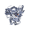 7tloC  2yooS S: Starting model for refinement C: citing same article ( |
|---|---|
| Similar structure data |
-Links
 PDBj
PDBj
- Assembly
| Deposited unit | 
| ||||||||
|---|---|---|---|---|---|---|---|---|---|
| 1 | 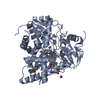
| ||||||||
| 2 | 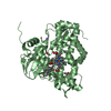
| ||||||||
| 3 | 
| ||||||||
| 4 | 
| ||||||||
| Unit cell |
|
-Components
-Protein , 1 types, 4 molecules ABCD
| #1: Protein | Mass: 45362.465 Da / Num. of mol.: 4 Source method: isolated from a genetically manipulated source Source: (gene. exp.) Mycobacterium ulcerans (bacteria) / Strain: Agy99 / Gene: cyp142A3, MUL_4077 / Production host: |
|---|
-Non-polymers , 5 types, 1670 molecules 
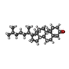


| #2: Chemical | ChemComp-HEM /  Mass: 616.487 Da / Num. of mol.: 4 Mass: 616.487 Da / Num. of mol.: 4Source method: isolated from a genetically manipulated source Formula: C34H32FeN4O4 / Feature type: SUBJECT OF INVESTIGATION #3: Chemical | ChemComp-K2B / (  Mass: 384.638 Da / Num. of mol.: 4 / Source method: obtained synthetically / Formula: C27H44O / Feature type: SUBJECT OF INVESTIGATION Mass: 384.638 Da / Num. of mol.: 4 / Source method: obtained synthetically / Formula: C27H44O / Feature type: SUBJECT OF INVESTIGATION#4: Chemical | ChemComp-NA /  Mass: 22.990 Da / Num. of mol.: 13 / Source method: obtained synthetically / Formula: Na Mass: 22.990 Da / Num. of mol.: 13 / Source method: obtained synthetically / Formula: Na#5: Chemical | ChemComp-ACT /  Mass: 59.044 Da / Num. of mol.: 7 / Source method: obtained synthetically / Formula: C2H3O2 Mass: 59.044 Da / Num. of mol.: 7 / Source method: obtained synthetically / Formula: C2H3O2#6: Water | ChemComp-HOH / | Mass: 18.015 Da / Num. of mol.: 1642 / Source method: isolated from a natural source / Formula: H2O |
|---|
-Details
| Has ligand of interest | Y |
|---|
-Experimental details
-Experiment
| Experiment | Method: X-RAY DIFFRACTION / Number of used crystals: 1 |
|---|
- Sample preparation
| Crystal | Density Matthews: 2.11 Å3/Da / Density % sol: 41.77 % |
|---|---|
| Crystal grow | Temperature: 288 K / Method: vapor diffusion, hanging drop Details: 0.2 M Sodium Acetate pH 5.5-6.0, 10% PEG 8000, 0.1 M Sodium cacodylate trihydrate PH range: 5.5-6.0 |
-Data collection
| Diffraction | Mean temperature: 100 K / Serial crystal experiment: N | ||||||||||||||||||||||||||||||
|---|---|---|---|---|---|---|---|---|---|---|---|---|---|---|---|---|---|---|---|---|---|---|---|---|---|---|---|---|---|---|---|
| Diffraction source | Source: SYNCHROTRON / Site: Australian Synchrotron / Beamline: MX2 / Wavelength: 0.95 Å | ||||||||||||||||||||||||||||||
| Detector | Type: ADSC QUANTUM 315r / Detector: CCD / Date: Apr 29, 2021 | ||||||||||||||||||||||||||||||
| Radiation | Protocol: SINGLE WAVELENGTH / Monochromatic (M) / Laue (L): M / Scattering type: x-ray | ||||||||||||||||||||||||||||||
| Radiation wavelength | Wavelength: 0.95 Å / Relative weight: 1 | ||||||||||||||||||||||||||||||
| Reflection | Resolution: 1.83→47.87 Å / Num. obs: 132141 / % possible obs: 98.6 % / Redundancy: 7.1 % / Biso Wilson estimate: 21.1 Å2 / CC1/2: 1 / Rmerge(I) obs: 0.052 / Rpim(I) all: 0.021 / Rrim(I) all: 0.056 / Net I/σ(I): 18.2 / Num. measured all: 935212 / Scaling rejects: 7 | ||||||||||||||||||||||||||||||
| Reflection shell | Diffraction-ID: 1
|
- Processing
| Software |
| ||||||||||||||||||||||||||||||||||||||||||||||||||||||||||||||||||||||||||||||||||||||||||
|---|---|---|---|---|---|---|---|---|---|---|---|---|---|---|---|---|---|---|---|---|---|---|---|---|---|---|---|---|---|---|---|---|---|---|---|---|---|---|---|---|---|---|---|---|---|---|---|---|---|---|---|---|---|---|---|---|---|---|---|---|---|---|---|---|---|---|---|---|---|---|---|---|---|---|---|---|---|---|---|---|---|---|---|---|---|---|---|---|---|---|---|
| Refinement | Method to determine structure: MOLECULAR REPLACEMENT Starting model: 2YOO Resolution: 1.83→40.807 Å / SU ML: 0.16 / Cross valid method: THROUGHOUT / σ(F): 1.36 / Phase error: 20.12 / Stereochemistry target values: ML
| ||||||||||||||||||||||||||||||||||||||||||||||||||||||||||||||||||||||||||||||||||||||||||
| Solvent computation | Shrinkage radii: 0.9 Å / VDW probe radii: 1.11 Å / Solvent model: FLAT BULK SOLVENT MODEL | ||||||||||||||||||||||||||||||||||||||||||||||||||||||||||||||||||||||||||||||||||||||||||
| Displacement parameters | Biso max: 83.26 Å2 / Biso mean: 25.2768 Å2 / Biso min: 8.92 Å2 | ||||||||||||||||||||||||||||||||||||||||||||||||||||||||||||||||||||||||||||||||||||||||||
| Refinement step | Cycle: final / Resolution: 1.83→40.807 Å
| ||||||||||||||||||||||||||||||||||||||||||||||||||||||||||||||||||||||||||||||||||||||||||
| Refine LS restraints |
| ||||||||||||||||||||||||||||||||||||||||||||||||||||||||||||||||||||||||||||||||||||||||||
| LS refinement shell | Refine-ID: X-RAY DIFFRACTION / Rfactor Rfree error: 0
|
