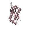
ジャーナル: Proc Natl Acad Sci U S A / 年: 2022 タイトル: The flagellar motor protein FliL forms a scaffold of circumferentially positioned rings required for stator activation. 著者: Shoichi Tachiyama / Kar L Chan / Xiaolin Liu / Skander Hathroubi / Briana Peterson / Mohammad F Khan / Karen M Ottemann / Jun Liu / Anna Roujeinikova / 要旨: The flagellar motor stator is an ion channel nanomachine that assembles as a ring of the MotAMotB units at the flagellar base. The role of accessory proteins required for stator assembly and ...The flagellar motor stator is an ion channel nanomachine that assembles as a ring of the MotAMotB units at the flagellar base. The role of accessory proteins required for stator assembly and activation remains largely enigmatic. Here, we show that one such assembly factor, the conserved protein FliL, forms an integral part of the flagellar motor in a position that colocalizes with the stator. Cryogenic electron tomography reconstructions of the intact motor in whole wild-type cells and cells lacking FliL revealed that the periplasmic domain of FliL (FliL-C) forms 18 circumferentially positioned rings integrated with the 18 MotAB units. FliL-C formed partial rings in the crystal, and the crystal structure-based full ring model was consistent with the shape of the rings observed in situ. Our data suggest that each FliL ring is coaxially sandwiched between the MotA ring and the dimeric periplasmic MotB moiety of the stator unit and that the central hole of the FliL ring has density that is consistent with the plug/linker region of MotB in its extended, active conformation. Significant structural similarities were found between FliL-C and stomatin/prohibitin/flotillin/HflK/C domains of scaffolding proteins, suggesting that FliL acts as a scaffold. The binding energy released upon association of FliL with the stator units could be used to power the release of the plug helices. The finding that isolated FliL-C forms stable partial rings provides an insight into the putative mechanism by which the FliL rings assemble around the stator units.
履歴
登録
2021年10月6日
登録サイト: RCSB / 処理サイト: RCSB
改定 1.0
2022年3月23日
Provider: repository / タイプ: Initial release
改定 1.1
2024年5月22日
Group: Data collection / カテゴリ: chem_comp_atom / chem_comp_bond
構造決定の手法: AB INITIO PHASING / 解像度: 2.1→42.76 Å / Cor.coef. Fo:Fc: 0.968 / Cor.coef. Fo:Fc free: 0.946 / SU B: 4.123 / SU ML: 0.11 / 交差検証法: THROUGHOUT / σ(F): 0 / ESU R: 0.158 / ESU R Free: 0.158 / 立体化学のターゲット値: MAXIMUM LIKELIHOOD 詳細: HYDROGENS HAVE BEEN ADDED IN THE RIDING POSITIONS U VALUES : REFINED INDIVIDUALLY
Rfactor
反射数
%反射
Selection details
Rfree
0.2329
439
4.8 %
RANDOM
Rwork
0.1793
-
-
-
obs
0.1817
8723
98.35 %
-
溶媒の処理
イオンプローブ半径: 0.8 Å / 減衰半径: 0.8 Å / VDWプローブ半径: 1.2 Å / 溶媒モデル: MASK
 ムービー
ムービー コントローラー


 データを開く
 基本情報
基本情報 要素
要素 キーワード
キーワード 機能・相同性情報
 Helicobacter pylori (ピロリ菌)
 X線回折 /
  データ登録者
 オーストラリア, 1件
  引用
引用
 構造の表示
構造の表示 ダウンロードとリンク
 その他のダウンロード

 PDBj
PDBj 集合体
集合体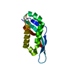
 分子量: 18.015 Da / 分子数: 61 / 由来タイプ: 天然 / 式: H2O
 試料調製
試料調製 解析
解析