[English] 日本語
Yorodumi
- PDB-7sgn: Crystal structure of periplasmic domain of Helicobacter pylori Fl... -

+
Open data


ID or keywords:

Loading...

-
Basic information

Entry
Database: PDB / ID: 7sgn
TitleCrystal structure of periplasmic domain of Helicobacter pylori FliL (residues 81 to 183) (crystal form A)
ComponentsFlagellar protein FliL
KeywordsMOTOR PROTEIN / Flagellar motor / FliL
Function / homologyFlagellar basal body-associated protein FliL / Flagellar basal body-associated protein FliL / bacterial-type flagellum basal body / bacterial-type flagellum-dependent cell motility / chemotaxis / plasma membrane / Flagellar protein FliL
Function and homology information
Biological speciesHelicobacter pylori (bacteria)
MethodX-RAY DIFFRACTION / SYNCHROTRON / MOLECULAR REPLACEMENT / Resolution: 2.8 Å
AuthorsChan, K.L. / Peterson, B. / Khan, M.F. / Roujeinikova, A.
Funding support Australia, 1items
OrganizationGrant numberCountry
Australian Research Council (ARC)DP210103056 Australia
Citation
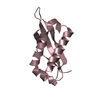
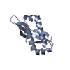
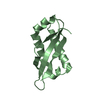
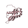
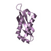
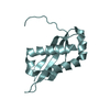
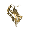
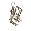
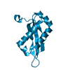
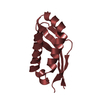
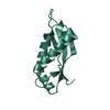
Journal: Proc Natl Acad Sci U S A / Year: 2022
Title: The flagellar motor protein FliL forms a scaffold of circumferentially positioned rings required for stator activation.
Authors: Shoichi Tachiyama / Kar L Chan / Xiaolin Liu / Skander Hathroubi / Briana Peterson / Mohammad F Khan / Karen M Ottemann / Jun Liu / Anna Roujeinikova /
Abstract: The flagellar motor stator is an ion channel nanomachine that assembles as a ring of the MotAMotB units at the flagellar base. The role of accessory proteins required for stator assembly and ...The flagellar motor stator is an ion channel nanomachine that assembles as a ring of the MotAMotB units at the flagellar base. The role of accessory proteins required for stator assembly and activation remains largely enigmatic. Here, we show that one such assembly factor, the conserved protein FliL, forms an integral part of the flagellar motor in a position that colocalizes with the stator. Cryogenic electron tomography reconstructions of the intact motor in whole wild-type cells and cells lacking FliL revealed that the periplasmic domain of FliL (FliL-C) forms 18 circumferentially positioned rings integrated with the 18 MotAB units. FliL-C formed partial rings in the crystal, and the crystal structure-based full ring model was consistent with the shape of the rings observed in situ. Our data suggest that each FliL ring is coaxially sandwiched between the MotA ring and the dimeric periplasmic MotB moiety of the stator unit and that the central hole of the FliL ring has density that is consistent with the plug/linker region of MotB in its extended, active conformation. Significant structural similarities were found between FliL-C and stomatin/prohibitin/flotillin/HflK/C domains of scaffolding proteins, suggesting that FliL acts as a scaffold. The binding energy released upon association of FliL with the stator units could be used to power the release of the plug helices. The finding that isolated FliL-C forms stable partial rings provides an insight into the putative mechanism by which the FliL rings assemble around the stator units.
History
DepositionOct 6, 2021Deposition site: RCSB / Processing site: RCSB
Revision 1.0Mar 23, 2022Provider: repository / Type: Initial release
Revision 1.1May 22, 2024Group: Data collection / Category: chem_comp_atom / chem_comp_bond

-
Structure visualization

Structure viewerMolecule:
MolmilJmol/JSmol

Downloads & links

-
Assembly

Deposited unit
A: Flagellar protein FliL
B: Flagellar protein FliL
C: Flagellar protein FliL
D: Flagellar protein FliL
E: Flagellar protein FliL
F: Flagellar protein FliL
G: Flagellar protein FliL
H: Flagellar protein FliL
I: Flagellar protein FliL
J: Flagellar protein FliL
K: Flagellar protein FliL
L: Flagellar protein FliL


Theoretical massNumber of molelcules
Total (without water)152,21512
Polymers152,21512
Non-polymers00
Water5,332296
1
A: Flagellar protein FliL


Theoretical massNumber of molelcules
Total (without water)12,6851
Polymers12,6851
Non-polymers00
Water181
TypeNameSymmetry operationNumber
identity operation1_555x,y,z1
MethodPISA
2
B: Flagellar protein FliL


Theoretical massNumber of molelcules
Total (without water)12,6851
Polymers12,6851
Non-polymers00
Water181
TypeNameSymmetry operationNumber
identity operation1_555x,y,z1
MethodPISA
3
C: Flagellar protein FliL


Theoretical massNumber of molelcules
Total (without water)12,6851
Polymers12,6851
Non-polymers00
Water181
TypeNameSymmetry operationNumber
identity operation1_555x,y,z1
MethodPISA
4
D: Flagellar protein FliL


Theoretical massNumber of molelcules
Total (without water)12,6851
Polymers12,6851
Non-polymers00
Water181
TypeNameSymmetry operationNumber
identity operation1_555x,y,z1
MethodPISA
5
E: Flagellar protein FliL


Theoretical massNumber of molelcules
Total (without water)12,6851
Polymers12,6851
Non-polymers00
Water181
TypeNameSymmetry operationNumber
identity operation1_555x,y,z1
MethodPISA
6
F: Flagellar protein FliL


Theoretical massNumber of molelcules
Total (without water)12,6851
Polymers12,6851
Non-polymers00
Water181
TypeNameSymmetry operationNumber
identity operation1_555x,y,z1
MethodPISA
7
G: Flagellar protein FliL


Theoretical massNumber of molelcules
Total (without water)12,6851
Polymers12,6851
Non-polymers00
Water181
TypeNameSymmetry operationNumber
identity operation1_555x,y,z1
MethodPISA
8
H: Flagellar protein FliL


Theoretical massNumber of molelcules
Total (without water)12,6851
Polymers12,6851
Non-polymers00
Water181
TypeNameSymmetry operationNumber
identity operation1_555x,y,z1
MethodPISA
9
I: Flagellar protein FliL


Theoretical massNumber of molelcules
Total (without water)12,6851
Polymers12,6851
Non-polymers00
Water181
TypeNameSymmetry operationNumber
identity operation1_555x,y,z1
MethodPISA
10
J: Flagellar protein FliL


Theoretical massNumber of molelcules
Total (without water)12,6851
Polymers12,6851
Non-polymers00
Water181
TypeNameSymmetry operationNumber
identity operation1_555x,y,z1
MethodPISA
11
K: Flagellar protein FliL


Theoretical massNumber of molelcules
Total (without water)12,6851
Polymers12,6851
Non-polymers00
Water181
TypeNameSymmetry operationNumber
identity operation1_555x,y,z1
MethodPISA
12
L: Flagellar protein FliL


Theoretical massNumber of molelcules
Total (without water)12,6851
Polymers12,6851
Non-polymers00
Water181
TypeNameSymmetry operationNumber
identity operation1_555x,y,z1
MethodPISA
Unit cell
Length a, b, c (Å)62.501, 82.550, 97.840
Angle α, β, γ (deg.)67.720, 83.390, 72.820
Int Tables number1
Space group name H-MP1

-
Components

#1: Protein
Flagellar protein FliL


Mass: 12684.601 Da / Num. of mol.: 12
Source method: isolated from a genetically manipulated source
Source: (gene. exp.) Helicobacter pylori (bacteria) / Gene: fliL, BGL67_04255 / Production host: Escherichia coli BL21(DE3) (bacteria) / References: UniProt: A0A293T1P9
#2: Water ChemComp-HOH / water


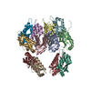
Mass: 18.015 Da / Num. of mol.: 296 / Source method: isolated from a natural source / Formula: H2O

-
Experimental details

-
Experiment

ExperimentMethod: X-RAY DIFFRACTION / Number of used crystals: 1

-
Sample preparation

CrystalDensity Matthews: 2.93 Å3/Da / Density % sol: 58.05 %
Crystal growTemperature: 298 K / Method: vapor diffusion, hanging drop
Details: Ammonium di-hydrogen phosphate and trisodium citrate

-
Data collection

DiffractionMean temperature: 100 K / Serial crystal experiment: N
Diffraction sourceSource: SYNCHROTRON / Site: Australian Synchrotron / Beamline: MX1 / Wavelength: 1 Å
DetectorType: ADSC QUANTUM 210r / Detector: CCD / Date: Jun 15, 2018
RadiationProtocol: SINGLE WAVELENGTH / Monochromatic (M) / Laue (L): M / Scattering type: x-ray
Radiation wavelengthWavelength: 1 Å / Relative weight: 1
ReflectionResolution: 2.8→54.16 Å / Num. obs: 41760 / % possible obs: 98 % / Redundancy: 2 % / Rmerge(I) obs: 0.103 / Net I/σ(I): 5.2
Reflection shellResolution: 2.8→2.91 Å / Rmerge(I) obs: 0.39 / Num. unique obs: 4676

-
Processing

Software
NameVersionClassification
PHENIX1.11.1_2575refinement
PDB_EXTRACT3.27data extraction
XDSdata reduction
Aimlessdata scaling
PHASERphasing
RefinementMethod to determine structure: MOLECULAR REPLACEMENT / Resolution: 2.8→46.902 Å / SU ML: 0.39 / Cross valid method: THROUGHOUT / σ(F): 2 / Phase error: 25.53 / Stereochemistry target values: ML
RfactorNum. reflection% reflection
Rfree0.2575 1905 4.56 %
Rwork0.1684 39843 -
obs0.1723 41748 98.09 %
Solvent computationShrinkage radii: 0.9 Å / VDW probe radii: 1.11 Å / Solvent model: FLAT BULK SOLVENT MODEL
Displacement parametersBiso max: 137.8 Å2 / Biso mean: 35.3539 Å2 / Biso min: 4.83 Å2
Refinement stepCycle: final / Resolution: 2.8→46.902 Å
ProteinNucleic acidLigandSolventTotal
Num. atoms9658 0 0 296 9954
Biso mean---32.35 -
Num. residues----1232
Refine LS restraints
Refine-IDTypeDev idealNumber
X-RAY DIFFRACTIONf_bond_d0.0089944
X-RAY DIFFRACTIONf_angle_d0.96513442
X-RAY DIFFRACTIONf_chiral_restr0.0561630
X-RAY DIFFRACTIONf_plane_restr0.0051700
X-RAY DIFFRACTIONf_dihedral_angle_d20.7163725
LS refinement shell

Refine-ID: X-RAY DIFFRACTION / Rfactor Rfree error: 0

Resolution (Å)Rfactor RfreeNum. reflection RfreeRfactor RworkNum. reflection Rwork% reflection obs (%)
2.8-2.870.30791520.2258279498
2.87-2.94760.28031350.2043284598
2.9476-3.03430.29011290.2134284098
3.0343-3.13230.32891600.2163283898
3.1323-3.24420.32511510.2018280098
3.2442-3.3740.3181270.1972286798
3.374-3.52760.28151390.1782281298
3.5276-3.71350.2781510.1753283498
3.7135-3.9460.23121240.164287898
3.946-4.25050.19411270.1364285398
4.2505-4.67790.23521300.1214287699
4.6779-5.3540.19851300.1282286699
5.354-6.74220.24551180.1767287699

+
About Yorodumi

-
News

-
Feb 9, 2022. New format data for meta-information of EMDB entries

New format data for meta-information of EMDB entries

  • Version 3 of the EMDB header file is now the official format.
  • The previous official version 1.9 will be removed from the archive.

Related info.:EMDB header

External links:wwPDB to switch to version 3 of the EMDB data model

-
Aug 12, 2020. Covid-19 info

Covid-19 info

URL: https://pdbjlvh1.pdbj.org/emnavi/covid19.php

New page: Covid-19 featured information page in EM Navigator.

Related info.:Covid-19 info / Mar 5, 2020. Novel coronavirus structure data

+
Mar 5, 2020. Novel coronavirus structure data

Novel coronavirus structure data

Related info.:Yorodumi Speices / Aug 12, 2020. Covid-19 info

External links:COVID-19 featured content - PDBj / Molecule of the Month (242):Coronavirus Proteases

+
Jan 31, 2019. EMDB accession codes are about to change! (news from PDBe EMDB page)

EMDB accession codes are about to change! (news from PDBe EMDB page)

  • The allocation of 4 digits for EMDB accession codes will soon come to an end. Whilst these codes will remain in use, new EMDB accession codes will include an additional digit and will expand incrementally as the available range of codes is exhausted. The current 4-digit format prefixed with “EMD-” (i.e. EMD-XXXX) will advance to a 5-digit format (i.e. EMD-XXXXX), and so on. It is currently estimated that the 4-digit codes will be depleted around Spring 2019, at which point the 5-digit format will come into force.
  • The EM Navigator/Yorodumi systems omit the EMD- prefix.

Related info.:Q: What is EMD? / ID/Accession-code notation in Yorodumi/EM Navigator

External links:EMDB Accession Codes are Changing Soon! / Contact to PDBj

+
Jul 12, 2017. Major update of PDB

Major update of PDB

  • wwPDB released updated PDB data conforming to the new PDBx/mmCIF dictionary.
  • This is a major update changing the version number from 4 to 5, and with Remediation, in which all the entries are updated.
  • In this update, many items about electron microscopy experimental information are reorganized (e.g. em_software).
  • Now, EM Navigator and Yorodumi are based on the updated data.

External links:wwPDB Remediation / Enriched Model Files Conforming to OneDep Data Standards Now Available in the PDB FTP Archive

-
Yorodumi

Thousand views of thousand structures

  • Yorodumi is a browser for structure data from EMDB, PDB, SASBDB, etc.
  • This page is also the successor to EM Navigator detail page, and also detail information page/front-end page for Omokage search.
  • The word "yorodu" (or yorozu) is an old Japanese word meaning "ten thousand". "mi" (miru) is to see.

Related info.:EMDB / PDB / SASBDB / Comparison of 3 databanks / Yorodumi Search / Aug 31, 2016. New EM Navigator & Yorodumi / Yorodumi Papers / Jmol/JSmol / Function and homology information / Changes in new EM Navigator and Yorodumi

Read more