 Movie
Movie Controller

[English] 日本語
 Yorodumi
- PDB-7sch: Cryo-EM structure of the human Exostosin-1 and Exostosin-2 heterodimer -
+ Open data

- Basic information
| Entry | Database: PDB / ID: 7sch | ||||||
|---|---|---|---|---|---|---|---|
| Title | Cryo-EM structure of the human Exostosin-1 and Exostosin-2 heterodimer | ||||||
 Components Components |
| ||||||
 Keywords Keywords | TRANSFERASE / exostosin1 / exostosin2 / glycosyltransferase / heparan sulfate | ||||||
| Function / homology |  Function and homology information Function and homology informationglucuronosyl-N-acetylglucosaminyl-proteoglycan 4-alpha-N-acetylglucosaminyltransferase / N-acetylglucosaminyl-proteoglycan 4-beta-glucuronosyltransferase / heparan sulfate N-acetylglucosaminyltransferase activity / glucuronosyl-N-acetylglucosaminyl-proteoglycan 4-alpha-N-acetylglucosaminyltransferase activity / N-acetylglucosaminyl-proteoglycan 4-beta-glucuronosyltransferase activity / UDP-N-acetylglucosamine transferase complex / glucuronosyltransferase activity / Defective EXT2 causes exostoses 2 / Defective EXT1 causes exostoses 1, TRPS2 and CHDS / glycosaminoglycan biosynthetic process ...glucuronosyl-N-acetylglucosaminyl-proteoglycan 4-alpha-N-acetylglucosaminyltransferase / N-acetylglucosaminyl-proteoglycan 4-beta-glucuronosyltransferase / heparan sulfate N-acetylglucosaminyltransferase activity / glucuronosyl-N-acetylglucosaminyl-proteoglycan 4-alpha-N-acetylglucosaminyltransferase activity / N-acetylglucosaminyl-proteoglycan 4-beta-glucuronosyltransferase activity / UDP-N-acetylglucosamine transferase complex / glucuronosyltransferase activity / Defective EXT2 causes exostoses 2 / Defective EXT1 causes exostoses 1, TRPS2 and CHDS / glycosaminoglycan biosynthetic process / HS-GAG biosynthesis / heparan sulfate proteoglycan biosynthetic process / acetylglucosaminyltransferase activity / polysaccharide biosynthetic process / glycosyltransferase activity / catalytic complex / ossification / protein heterodimerization activity / Golgi membrane / endoplasmic reticulum membrane / Golgi apparatus / endoplasmic reticulum / protein homodimerization activity / extracellular exosome / membrane / metal ion binding Similarity search - Function | ||||||
| Biological species |  Homo sapiens (human) Homo sapiens (human) | ||||||
| Method | ELECTRON MICROSCOPY / single particle reconstruction / cryo EM / Resolution: 3.1 Å | ||||||
 Authors Authors | Li, H. / Li, H. | ||||||
| Funding support |  United States, 1items United States, 1items
| ||||||
 Citation Citation | Journal: Nat Chem Biol / Year: 2023 Title: Structural basis for heparan sulfate co-polymerase action by the EXT1-2 complex. Authors: Hua Li / Digantkumar Chapla / Robert A Amos / Annapoorani Ramiah / Kelley W Moremen / Huilin Li / Abstract: Heparan sulfate (HS) proteoglycans are extended (-GlcAβ1,4GlcNAcα1,4-) co-polymers containing decorations of sulfation and epimerization that are linked to cell surface and extracellular matrix ...Heparan sulfate (HS) proteoglycans are extended (-GlcAβ1,4GlcNAcα1,4-) co-polymers containing decorations of sulfation and epimerization that are linked to cell surface and extracellular matrix proteins. In mammals, HS repeat units are extended by an obligate heterocomplex of two exostosin family members, EXT1 and EXT2, where each protein monomer contains distinct GT47 (GT-B fold) and GT64 (GT-A fold) glycosyltransferase domains. In this study, we generated human EXT1-EXT2 (EXT1-2) as a functional heterocomplex and determined its structure in the presence of bound donor and acceptor substrates. Structural data and enzyme activity of catalytic site mutants demonstrate that only two of the four glycosyltransferase domains are major contributors to co-polymer syntheses: the EXT1 GT-B fold β1,4GlcA transferase domain and the EXT2 GT-A fold α1,4GlcNAc transferase domain. The two catalytic sites are over 90 Å apart, indicating that HS is synthesized by a dissociative process that involves a single catalytic site on each monomer. | ||||||
| History |
|
- Structure visualization
| Structure viewer | Molecule: MolmilJmol/JSmol |
|---|
- Downloads & links
-Download
| PDBx/mmCIF format | 7sch.cif.gz | 232 KB | Display | PDBx/mmCIF format |
|---|---|---|---|---|
| PDB format | pdb7sch.ent.gz | 177.5 KB | Display | PDB format |
| PDBx/mmJSON format | 7sch.json.gz | Tree view | PDBx/mmJSON format | |
| Others |  Other downloads Other downloads |
-Validation report
| Arichive directory | https://data.pdbj.org/pub/pdb/validation_reports/sc/7schftp://data.pdbj.org/pub/pdb/validation_reports/sc/7sch | HTTPS FTP |
|---|
-Related structure data
| Related structure data |  25035MC  7scjC  7sckC  7uqxC  7uqyC M: map data used to model this data C: citing same article ( |
|---|---|
| Similar structure data |


-Links
 PDBj
PDBj
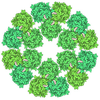
- Assembly
| Deposited unit | 
|
|---|---|
| 1 |
|
-Components
| #1: Protein | Mass: 83404.023 Da / Num. of mol.: 1 Source method: isolated from a genetically manipulated source Source: (gene. exp.) Homo sapiens (human) / Gene: EXT1 / Production host: Mammalia (mammals)References: UniProt: Q16394, glucuronosyl-N-acetylglucosaminyl-proteoglycan 4-alpha-N-acetylglucosaminyltransferase, N-acetylglucosaminyl-proteoglycan 4-beta-glucuronosyltransferase |
|---|---|
| #2: Protein | Mass: 77238.031 Da / Num. of mol.: 1 Source method: isolated from a genetically manipulated source Source: (gene. exp.) Homo sapiens (human) / Gene: EXT2 / Production host: Mammalia (mammals)References: UniProt: Q93063, glucuronosyl-N-acetylglucosaminyl-proteoglycan 4-alpha-N-acetylglucosaminyltransferase, N-acetylglucosaminyl-proteoglycan 4-beta-glucuronosyltransferase |
| #3: Polysaccharide | beta-D-mannopyranose-(1-4)-2-acetamido-2-deoxy-beta-D-glucopyranose-(1-4)-2-acetamido-2-deoxy-beta- ...beta-D-mannopyranose-(1-4)-2-acetamido-2-deoxy-beta-D-glucopyranose-(1-4)-2-acetamido-2-deoxy-beta-D-glucopyranose Source method: isolated from a genetically manipulated source |
| Has ligand of interest | Y |
| Has protein modification | Y |
-Experimental details
-Experiment
| Experiment | Method: ELECTRON MICROSCOPY |
|---|---|
| EM experiment | Aggregation state: PARTICLE / 3D reconstruction method: single particle reconstruction |
- Sample preparation
| Component | Name: hEXT1/2 / Type: COMPLEX / Entity ID: #1-#2 / Source: RECOMBINANT | |||||||||||||||
|---|---|---|---|---|---|---|---|---|---|---|---|---|---|---|---|---|
| Source (natural) | Organism: Homo sapiens (human) | |||||||||||||||
| Source (recombinant) | Organism: Mammalia (mammals) | |||||||||||||||
| Buffer solution | pH: 7 | |||||||||||||||
| Buffer component |
| |||||||||||||||
| Specimen | Conc.: 0.3 mg/ml / Embedding applied: NO / Shadowing applied: NO / Staining applied: NO / Vitrification applied: YES | |||||||||||||||
| Specimen support | Grid material: GOLD / Grid mesh size: 300 divisions/in. / Grid type: Quantifoil R2/1 | |||||||||||||||
| Vitrification | Instrument: FEI VITROBOT MARK IV / Cryogen name: ETHANE / Humidity: 95 % / Chamber temperature: 299 K |
- Electron microscopy imaging
| Experimental equipment |  Model: Titan Krios / Image courtesy: FEI Company |
|---|---|
| Microscopy | Model: FEI TITAN KRIOS |
| Electron gun | Electron source:  FIELD EMISSION GUN / Accelerating voltage: 300 kV / Illumination mode: OTHER FIELD EMISSION GUN / Accelerating voltage: 300 kV / Illumination mode: OTHER |
| Electron lens | Mode: BRIGHT FIELD / Nominal magnification: 105000 X / Cs: 2.7 mm / C2 aperture diameter: 70 µm / Alignment procedure: COMA FREE |
| Specimen holder | Cryogen: NITROGEN / Specimen holder model: FEI TITAN KRIOS AUTOGRID HOLDER / Temperature (max): 193 K / Temperature (min): 193 K / Residual tilt: 0.05 mradians |
| Image recording | Average exposure time: 1.5 sec. / Electron dose: 66.8 e/Å2 / Film or detector model: GATAN K3 (6k x 4k) / Num. of grids imaged: 1 / Num. of real images: 2980 |
| Image scans | Width: 5760 / Height: 4092 |
- Processing
| EM software |
| ||||||||||||||||||||
|---|---|---|---|---|---|---|---|---|---|---|---|---|---|---|---|---|---|---|---|---|---|
| CTF correction | Type: PHASE FLIPPING AND AMPLITUDE CORRECTION | ||||||||||||||||||||
| Particle selection | Num. of particles selected: 5186047 | ||||||||||||||||||||
| Symmetry | Point symmetry: C1 (asymmetric) | ||||||||||||||||||||
| 3D reconstruction | Resolution: 3.1 Å / Resolution method: FSC 0.143 CUT-OFF / Num. of particles: 120216 / Symmetry type: POINT | ||||||||||||||||||||
| Atomic model building | Protocol: AB INITIO MODEL |
