 Movie
Movie Controller


+ Open data

- Basic information
| Entry | Database: PDB / ID: 7sbm | |||||||||
|---|---|---|---|---|---|---|---|---|---|---|
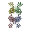
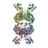
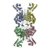
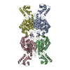
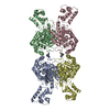
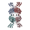
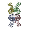
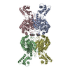
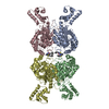
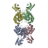
| Title | Human glutaminase C (Y466W) with L-Gln, open conformation | |||||||||
 Components Components | Isoform 3 of Glutaminase kidney isoform, mitochondrial | |||||||||
 Keywords Keywords | HYDROLASE / Complex / Oligomer / Cancer metabolism / Mitochondrial metabolism | |||||||||
| Function / homology |  Function and homology information Function and homology informationL-glutamine catabolic process / regulation of respiratory gaseous exchange by nervous system process / glutaminase / intracellular glutamate homeostasis / Glutamate and glutamine metabolism / Glutamate Neurotransmitter Release Cycle / : / glutaminase activity / suckling behavior / TP53 Regulates Metabolic Genes ...L-glutamine catabolic process / regulation of respiratory gaseous exchange by nervous system process / glutaminase / intracellular glutamate homeostasis / Glutamate and glutamine metabolism / Glutamate Neurotransmitter Release Cycle / : / glutaminase activity / suckling behavior / TP53 Regulates Metabolic Genes / protein homotetramerization / chemical synaptic transmission / mitochondrial matrix / synapse / mitochondrion / cytosol Similarity search - Function | |||||||||
| Biological species |  Homo sapiens (human) Homo sapiens (human) | |||||||||
| Method |  X-RAY DIFFRACTION / SYNCHROTRON / MOLECULAR REPLACEMENT / Resolution: 2.8 Å X-RAY DIFFRACTION / SYNCHROTRON / MOLECULAR REPLACEMENT / Resolution: 2.8 Å | |||||||||
 Authors Authors | Nguyen, T.-T.T. / Cerione, R.A. | |||||||||
| Funding support |  United States, 2items United States, 2items
| |||||||||
 Citation Citation | Journal: J.Biol.Chem. / Year: 2022 Title: High-resolution structures of mitochondrial glutaminase C tetramers indicate conformational changes upon phosphate binding. Authors: Nguyen, T.T. / Ramachandran, S. / Hill, M.J. / Cerione, R.A. | |||||||||
| History |
|
- Structure visualization
| Structure viewer | Molecule: MolmilJmol/JSmol |
|---|
- Downloads & links
-Download
| PDBx/mmCIF format | 7sbm.cif.gz | 747.8 KB | Display | PDBx/mmCIF format |
|---|---|---|---|---|
| PDB format | pdb7sbm.ent.gz | 515 KB | Display | PDB format |
| PDBx/mmJSON format | 7sbm.json.gz | Tree view | PDBx/mmJSON format | |
| Others |  Other downloads Other downloads |
-Validation report
| Arichive directory | https://data.pdbj.org/pub/pdb/validation_reports/sb/7sbmftp://data.pdbj.org/pub/pdb/validation_reports/sb/7sbm | HTTPS FTP |
|---|
-Related structure data
| Related structure data | 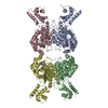 7sbnC 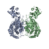 5d3oS S: Starting model for refinement C: citing same article ( |
|---|---|
| Similar structure data |


-Links
 PDBj
PDBj
- Assembly
| Deposited unit | 
| |||||||||||||||||||||||||||||||||||||||||||||||||||||||||||||||||||||||||||||||||||
|---|---|---|---|---|---|---|---|---|---|---|---|---|---|---|---|---|---|---|---|---|---|---|---|---|---|---|---|---|---|---|---|---|---|---|---|---|---|---|---|---|---|---|---|---|---|---|---|---|---|---|---|---|---|---|---|---|---|---|---|---|---|---|---|---|---|---|---|---|---|---|---|---|---|---|---|---|---|---|---|---|---|---|---|---|
| 1 | 
| |||||||||||||||||||||||||||||||||||||||||||||||||||||||||||||||||||||||||||||||||||
| Unit cell |
| |||||||||||||||||||||||||||||||||||||||||||||||||||||||||||||||||||||||||||||||||||
| Noncrystallographic symmetry (NCS) | NCS domain:
NCS domain segments: Ens-ID: 1
|