 Movie
Movie Controller

[English] 日本語
 Yorodumi
- PDB-7san: Crystal structure of human hypoxanthine guanine phosphoribzosyltr... -
+ Open data

- Basic information
| Entry | Database: PDB / ID: 7san | ||||||
|---|---|---|---|---|---|---|---|
| Title | Crystal structure of human hypoxanthine guanine phosphoribzosyltransferase in complex with (4S,7S)-7-hydroxy-4-((guanin-9-yl)methyl)-2,5-dioxaheptan-1,7-diphosphonate | ||||||
 Components Components | Hypoxanthine-guanine phosphoribosyltransferase | ||||||
 Keywords Keywords | TRANSFERASE/TRANSFERASE INHIBITOR / inhibitor / complex / phosphonate / purine / TRANSFERASE / TRANSFERASE-TRANSFERASE INHIBITOR complex | ||||||
| Function / homology |  Function and homology information Function and homology informationDefective HPRT1 disrupts guanine and hypoxanthine salvage / positive regulation of dopamine metabolic process / GMP catabolic process / hypoxanthine phosphoribosyltransferase / guanine phosphoribosyltransferase activity / guanine salvage / hypoxanthine metabolic process / hypoxanthine salvage / IMP metabolic process / hypoxanthine phosphoribosyltransferase activity ...Defective HPRT1 disrupts guanine and hypoxanthine salvage / positive regulation of dopamine metabolic process / GMP catabolic process / hypoxanthine phosphoribosyltransferase / guanine phosphoribosyltransferase activity / guanine salvage / hypoxanthine metabolic process / hypoxanthine salvage / IMP metabolic process / hypoxanthine phosphoribosyltransferase activity / GMP salvage / Purine salvage / IMP salvage / AMP salvage / purine nucleotide biosynthetic process / Azathioprine ADME / purine ribonucleoside salvage / protein homotetramerization / nucleotide binding / magnesium ion binding / extracellular exosome / identical protein binding / cytosol / cytoplasm Similarity search - Function | ||||||
| Biological species |  Homo sapiens (human) Homo sapiens (human) | ||||||
| Method |  X-RAY DIFFRACTION / SYNCHROTRON / MOLECULAR REPLACEMENT / Resolution: 2.58155523622 Å X-RAY DIFFRACTION / SYNCHROTRON / MOLECULAR REPLACEMENT / Resolution: 2.58155523622 Å | ||||||
 Authors Authors | Guddat, L.W. / Keough, D.T. | ||||||
| Funding support |  Australia, 1items Australia, 1items
| ||||||
 Citation Citation | Journal: J.Med.Chem. / Year: 2022 Title: Stereo-Defined Acyclic Nucleoside Phosphonates are Selective and Potent Inhibitors of Parasite 6-Oxopurine Phosphoribosyltransferases. Authors: Klejch, T. / Keough, D.T. / King, G. / Dolezelova, E. / Cesnek, M. / Budesinsky, M. / Zikova, A. / Janeba, Z. / Guddat, L.W. / Hockova, D. | ||||||
| History |
|
- Structure visualization
| Structure viewer | Molecule: MolmilJmol/JSmol |
|---|
- Downloads & links
-Download
| PDBx/mmCIF format | 7san.cif.gz | 184.9 KB | Display | PDBx/mmCIF format |
|---|---|---|---|---|
| PDB format | pdb7san.ent.gz | 134.9 KB | Display | PDB format |
| PDBx/mmJSON format | 7san.json.gz | Tree view | PDBx/mmJSON format | |
| Others |  Other downloads Other downloads |
-Validation report
| Arichive directory | https://data.pdbj.org/pub/pdb/validation_reports/sa/7sanftp://data.pdbj.org/pub/pdb/validation_reports/sa/7san | HTTPS FTP |
|---|
-Related structure data
| Related structure data |  7sb7C  7scrC 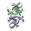 3gepS S: Starting model for refinement C: citing same article ( |
|---|---|
| Similar structure data |
-Links
 PDBj
PDBj
- Assembly
| Deposited unit | 
| ||||||||||||
|---|---|---|---|---|---|---|---|---|---|---|---|---|---|
| 1 |
| ||||||||||||
| Unit cell |
|
-Components
| #1: Protein | Mass: 24385.021 Da / Num. of mol.: 4 / Mutation: C22A, C105A, C205A Source method: isolated from a genetically manipulated source Source: (gene. exp.) Homo sapiens (human) / Gene: HPRT1, HPRT / Production host:  References: UniProt: P00492, hypoxanthine phosphoribosyltransferase #2: Chemical | ChemComp-8QI / ({(   Mass: 443.244 Da / Num. of mol.: 4 / Source method: obtained synthetically / Formula: C11H19N5O10P2 / Feature type: SUBJECT OF INVESTIGATION Mass: 443.244 Da / Num. of mol.: 4 / Source method: obtained synthetically / Formula: C11H19N5O10P2 / Feature type: SUBJECT OF INVESTIGATION#3: Water | ChemComp-HOH / |  Mass: 18.015 Da / Num. of mol.: 60 / Source method: isolated from a natural source / Formula: H2O Mass: 18.015 Da / Num. of mol.: 60 / Source method: isolated from a natural source / Formula: H2OHas ligand of interest | Y | |
|---|
-Experimental details
-Experiment
| Experiment | Method: X-RAY DIFFRACTION / Number of used crystals: 1 |
|---|
- Sample preparation
| Crystal | Density Matthews: 2.34 Å3/Da / Density % sol: 47.52 % |
|---|---|
| Crystal grow | Temperature: 293 K / Method: vapor diffusion, hanging drop / pH: 7.4 Details: 0.1 M Bis-tris propane, pH 7.4, 0.1 M sodium citrate, 17.5% PEG 3350 Temp details: INCUBATOR |
-Data collection
| Diffraction | Mean temperature: 100 K / Serial crystal experiment: N |
|---|---|
| Diffraction source | Source: SYNCHROTRON / Site: Australian Synchrotron / Beamline: MX1 / Wavelength: 0.987 Å |
| Detector | Type: DECTRIS PILATUS 2M / Detector: PIXEL / Date: Jul 1, 2020 |
| Radiation | Protocol: SINGLE WAVELENGTH / Monochromatic (M) / Laue (L): M / Scattering type: x-ray |
| Radiation wavelength | Wavelength: 0.987 Å / Relative weight: 1 |
| Reflection | Resolution: 2.58→49.17 Å / Num. obs: 29515 / % possible obs: 99.6 % / Redundancy: 11.2 % / Biso Wilson estimate: 50.4065251836 Å2 / CC1/2: 0.97 / Net I/σ(I): 12.66 |
| Reflection shell | Resolution: 2.58→2.67 Å / Redundancy: 7.5 % / Num. unique obs: 2806 / CC1/2: 0.9 / % possible all: 96.19 |
- Processing
| Software |
| |||||||||||||||||||||||||||||||||||||||||||||||||||||||||||||||||||||||||||||||||||||||||||||||||||||||||
|---|---|---|---|---|---|---|---|---|---|---|---|---|---|---|---|---|---|---|---|---|---|---|---|---|---|---|---|---|---|---|---|---|---|---|---|---|---|---|---|---|---|---|---|---|---|---|---|---|---|---|---|---|---|---|---|---|---|---|---|---|---|---|---|---|---|---|---|---|---|---|---|---|---|---|---|---|---|---|---|---|---|---|---|---|---|---|---|---|---|---|---|---|---|---|---|---|---|---|---|---|---|---|---|---|---|---|
| Refinement | Method to determine structure: MOLECULAR REPLACEMENT Starting model: 3GEP Resolution: 2.58155523622→49.1672764131 Å / SU ML: 0.337542685968 / Cross valid method: FREE R-VALUE / σ(F): 1.34356605534 / Phase error: 29.7639423357
| |||||||||||||||||||||||||||||||||||||||||||||||||||||||||||||||||||||||||||||||||||||||||||||||||||||||||
| Solvent computation | Shrinkage radii: 0.9 Å / VDW probe radii: 1.11 Å / Solvent model: FLAT BULK SOLVENT MODEL | |||||||||||||||||||||||||||||||||||||||||||||||||||||||||||||||||||||||||||||||||||||||||||||||||||||||||
| Displacement parameters | Biso mean: 51.9478388429 Å2 | |||||||||||||||||||||||||||||||||||||||||||||||||||||||||||||||||||||||||||||||||||||||||||||||||||||||||
| Refinement step | Cycle: LAST / Resolution: 2.58155523622→49.1672764131 Å
| |||||||||||||||||||||||||||||||||||||||||||||||||||||||||||||||||||||||||||||||||||||||||||||||||||||||||
| Refine LS restraints |
| |||||||||||||||||||||||||||||||||||||||||||||||||||||||||||||||||||||||||||||||||||||||||||||||||||||||||
| LS refinement shell |
|
