 Movie
Movie Controller


+ Open data

- Basic information
| Entry | Database: PDB / ID: 7sa8 | |||||||||
|---|---|---|---|---|---|---|---|---|---|---|
| Title | Crystal Structure of the periplasmic lyase AlgL K66A Mutant | |||||||||
 Components Components | Alginate lyase | |||||||||
 Keywords Keywords | LYASE / Alginate lyase | |||||||||
| Function / homology |  Function and homology information Function and homology informationalginic acid catabolic process / poly(beta-D-mannuronate) lyase activity / periplasmic space Similarity search - Function | |||||||||
| Biological species |   Pseudomonas aeruginosa (bacteria) Pseudomonas aeruginosa (bacteria) | |||||||||
| Method |  X-RAY DIFFRACTION / MOLECULAR REPLACEMENT / Resolution: 2.5 Å X-RAY DIFFRACTION / MOLECULAR REPLACEMENT / Resolution: 2.5 Å | |||||||||
 Authors Authors | Gheorghita, A.A. / Pfoh, R. / Wong, S.S.Y. / Howell, P.L. | |||||||||
| Funding support |  Canada, 2items Canada, 2items
| |||||||||
 Citation Citation | Journal: J.Biol.Chem. / Year: 2022 Title: The Pseudomonas aeruginosa homeostasis enzyme AlgL clears the periplasmic space of accumulated alginate during polymer biosynthesis. Authors: Gheorghita, A.A. / Wolfram, F. / Whitfield, G.B. / Jacobs, H.M. / Pfoh, R. / Wong, S.S.Y. / Guitor, A.K. / Goodyear, M.C. / Berezuk, A.M. / Khursigara, C.M. / Parsek, M.R. / Howell, P.L. | |||||||||
| History |
|
- Structure visualization
| Structure viewer | Molecule: MolmilJmol/JSmol |
|---|
- Downloads & links
-Download
| PDBx/mmCIF format | 7sa8.cif.gz | 128.6 KB | Display | PDBx/mmCIF format |
|---|---|---|---|---|
| PDB format | pdb7sa8.ent.gz | 95.4 KB | Display | PDB format |
| PDBx/mmJSON format | 7sa8.json.gz | Tree view | PDBx/mmJSON format | |
| Others |  Other downloads Other downloads |
-Validation report
| Arichive directory | https://data.pdbj.org/pub/pdb/validation_reports/sa/7sa8ftp://data.pdbj.org/pub/pdb/validation_reports/sa/7sa8 | HTTPS FTP |
|---|
-Related structure data
| Related structure data |  4ozvSC  4ozwC S: Starting model for refinement C: citing same article ( |
|---|---|
| Similar structure data |


-Links
 PDBj
PDBj- Assembly
| Deposited unit | 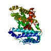
| ||||||||
|---|---|---|---|---|---|---|---|---|---|
| 1 |
| ||||||||
| Unit cell |
|
-Components
| #1: Protein | Mass: 39820.723 Da / Num. of mol.: 1 Source method: isolated from a genetically manipulated source Source: (gene. exp.) Pseudomonas aeruginosa (bacteria) / Gene: algL / Production host: |
|---|---|
| #2: Water | ChemComp-HOH /  Mass: 18.015 Da / Num. of mol.: 38 / Source method: isolated from a natural source / Formula: H2O Mass: 18.015 Da / Num. of mol.: 38 / Source method: isolated from a natural source / Formula: H2O |
| Has protein modification | Y |
-Experimental details
-Experiment
| Experiment | Method: X-RAY DIFFRACTION / Number of used crystals: 1 |
|---|
- Sample preparation
| Crystal | Density Matthews: 1.9 Å3/Da / Density % sol: 35.16 % |
|---|---|
| Crystal grow | Temperature: 293 K / Method: vapor diffusion, hanging drop Details: 0.275 M K2SO4, 19% (w/v) PEG 3350, and 0.1 M HEPES pH 6.9. Crystals were cryoprotected by soaking for 10 min with 2 mM mannuronate tetrasaccharide and 20% (v/v) PEG 4000. |
-Data collection
| Diffraction | Mean temperature: 100 K / Serial crystal experiment: N |
|---|---|
| Diffraction source | Source: SEALED TUBE / Type: BRUKER D8 QUEST / Wavelength: 1.5406 Å |
| Detector | Type: BRUKER PHOTON 100 / Detector: CMOS / Date: May 13, 2015 |
| Radiation | Monochromator: M / Protocol: SINGLE WAVELENGTH / Monochromatic (M) / Laue (L): M / Scattering type: x-ray |
| Radiation wavelength | Wavelength: 1.5406 Å / Relative weight: 1 |
| Reflection | Resolution: 2.5→20.305 Å / Num. obs: 10903 / % possible obs: 99.3 % / Redundancy: 10.3 % / Rmerge(I) obs: 0.098 / Net I/σ(I): 16 |
| Reflection shell | Resolution: 2.5→2.6 Å / Rmerge(I) obs: 0.435 / Num. unique obs: 1196 / % possible all: 100 |
- Processing
| Software |
| |||||||||||||||||||||||||||||||||||||||||||||||||||||||||||||||||||||||||||||||||||||||||||||||||||||||||||||||||||||||||||||||||||||||||||||||||||||||||||
|---|---|---|---|---|---|---|---|---|---|---|---|---|---|---|---|---|---|---|---|---|---|---|---|---|---|---|---|---|---|---|---|---|---|---|---|---|---|---|---|---|---|---|---|---|---|---|---|---|---|---|---|---|---|---|---|---|---|---|---|---|---|---|---|---|---|---|---|---|---|---|---|---|---|---|---|---|---|---|---|---|---|---|---|---|---|---|---|---|---|---|---|---|---|---|---|---|---|---|---|---|---|---|---|---|---|---|---|---|---|---|---|---|---|---|---|---|---|---|---|---|---|---|---|---|---|---|---|---|---|---|---|---|---|---|---|---|---|---|---|---|---|---|---|---|---|---|---|---|---|---|---|---|---|---|---|---|
| Refinement | Method to determine structure: MOLECULAR REPLACEMENT Starting model: 4OZV Resolution: 2.5→20.305 Å / Cor.coef. Fo:Fc: 0.912 / Cor.coef. Fo:Fc free: 0.878 / SU B: 29.256 / SU ML: 0.295 / Cross valid method: THROUGHOUT / ESU R: 1.643 / ESU R Free: 0.337 Details: Hydrogens have been added in their riding positions
| |||||||||||||||||||||||||||||||||||||||||||||||||||||||||||||||||||||||||||||||||||||||||||||||||||||||||||||||||||||||||||||||||||||||||||||||||||||||||||
| Solvent computation | Ion probe radii: 0.8 Å / Shrinkage radii: 0.8 Å / VDW probe radii: 1.2 Å / Solvent model: MASK BULK SOLVENT | |||||||||||||||||||||||||||||||||||||||||||||||||||||||||||||||||||||||||||||||||||||||||||||||||||||||||||||||||||||||||||||||||||||||||||||||||||||||||||
| Displacement parameters | Biso mean: 39.375 Å2
| |||||||||||||||||||||||||||||||||||||||||||||||||||||||||||||||||||||||||||||||||||||||||||||||||||||||||||||||||||||||||||||||||||||||||||||||||||||||||||
| Refinement step | Cycle: LAST / Resolution: 2.5→20.305 Å
| |||||||||||||||||||||||||||||||||||||||||||||||||||||||||||||||||||||||||||||||||||||||||||||||||||||||||||||||||||||||||||||||||||||||||||||||||||||||||||
| Refine LS restraints |
| |||||||||||||||||||||||||||||||||||||||||||||||||||||||||||||||||||||||||||||||||||||||||||||||||||||||||||||||||||||||||||||||||||||||||||||||||||||||||||
| LS refinement shell |
| |||||||||||||||||||||||||||||||||||||||||||||||||||||||||||||||||||||||||||||||||||||||||||||||||||||||||||||||||||||||||||||||||||||||||||||||||||||||||||
| Refinement TLS params. | Method: refined / Origin x: 9.039 Å / Origin y: 82.556 Å / Origin z: 19.965 Å
| |||||||||||||||||||||||||||||||||||||||||||||||||||||||||||||||||||||||||||||||||||||||||||||||||||||||||||||||||||||||||||||||||||||||||||||||||||||||||||
| Refinement TLS group | Selection: ALL |
