 Movie
Movie Controller

[English] 日本語
 Yorodumi
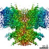
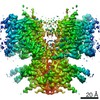
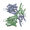
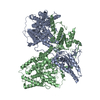
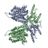
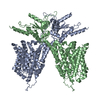
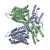
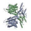
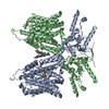
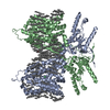
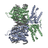
- PDB-7s9b: Cryo-EM Structure of dolphin Prestin: Sensor Down I (Expanded) state -
+ Open data

- Basic information
| Entry | Database: PDB / ID: 7s9b | ||||||
|---|---|---|---|---|---|---|---|
| Title | Cryo-EM Structure of dolphin Prestin: Sensor Down I (Expanded) state | ||||||
 Components Components | Prestin | ||||||
 Keywords Keywords | MOTOR PROTEIN / Outer hair cells / electromotility / mechanotransduction / hearing / deafness / frequency sensation / echolocation / SLC26 / SLC26A5 | ||||||
| Function / homology |  Function and homology information Function and homology informationcochlear outer hair cell electromotile response / secondary active sulfate transmembrane transporter activity / sensory perception of sound / regulation of cell shape / plasma membrane Similarity search - Function | ||||||
| Biological species |  | ||||||
| Method | ELECTRON MICROSCOPY / single particle reconstruction / cryo EM / Resolution: 4.2 Å | ||||||
 Authors Authors | Bavi, N. / Clark, M.D. / Contreras, G.F. / Shen, R. / Reddy, B.G. / Milewski, W. / Perozo, E. | ||||||
| Funding support |  United States, 1items United States, 1items
| ||||||
 Citation Citation | Journal: Nature / Year: 2021 Title: The conformational cycle of prestin underlies outer-hair cell electromotility. Authors: Navid Bavi / Michael David Clark / Gustavo F Contreras / Rong Shen / Bharat G Reddy / Wieslawa Milewski / Eduardo Perozo / Abstract: The voltage-dependent motor protein prestin (also known as SLC26A5) is responsible for the electromotive behaviour of outer-hair cells and underlies the cochlear amplifier. Knockout or impairment of ...The voltage-dependent motor protein prestin (also known as SLC26A5) is responsible for the electromotive behaviour of outer-hair cells and underlies the cochlear amplifier. Knockout or impairment of prestin causes severe hearing loss. Despite the key role of prestin in hearing, the mechanism by which mammalian prestin senses voltage and transduces it into cellular-scale movements (electromotility) is poorly understood. Here we determined the structure of dolphin prestin in six distinct states using single-particle cryo-electron microscopy. Our structural and functional data suggest that prestin adopts a unique and complex set of states, tunable by the identity of bound anions (Cl or SO). Salicylate, a drug that can cause reversible hearing loss, competes for the anion-binding site of prestin, and inhibits its function by immobilizing prestin in a new conformation. Our data suggest that the bound anion together with its coordinating charged residues and helical dipole act as a dynamic voltage sensor. An analysis of all of the anion-dependent conformations reveals how structural rearrangements in the voltage sensor are coupled to conformational transitions at the protein-membrane interface, suggesting a previously undescribed mechanism of area expansion. Visualization of the electromotility cycle of prestin distinguishes the protein from the closely related SLC26 anion transporters, highlighting the basis for evolutionary specialization of the mammalian cochlear amplifier at a high resolution. | ||||||
| History |
|
- Structure visualization
| Movie |
Movie viewer |
|---|---|
| Structure viewer | Molecule: MolmilJmol/JSmol |
- Downloads & links
-Download
| PDBx/mmCIF format | 7s9b.cif.gz | 234.1 KB | Display | PDBx/mmCIF format |
|---|---|---|---|---|
| PDB format | pdb7s9b.ent.gz | 190.3 KB | Display | PDB format |
| PDBx/mmJSON format | 7s9b.json.gz | Tree view | PDBx/mmJSON format | |
| Others |  Other downloads Other downloads |
-Validation report
| Arichive directory | https://data.pdbj.org/pub/pdb/validation_reports/s9/7s9bftp://data.pdbj.org/pub/pdb/validation_reports/s9/7s9b | HTTPS FTP |
|---|
-Related structure data
| Related structure data |  24931MC  7s8xC  7s9aC  7s9cC  7s9dC  7s9eC M: map data used to model this data C: citing same article ( |
|---|---|
| Similar structure data |


-Links
 PDBj
PDBj
- Assembly
| Deposited unit | 
|
|---|---|
| 1 |
|
-Components
| #1: Protein | Mass: 80973.750 Da / Num. of mol.: 2 Source method: isolated from a genetically manipulated source Source: (gene. exp.) Gene: SLC26A5 / Cell line (production host): HEK293 / Production host:  Homo sapiens (human) / References: UniProt: D7PC76 Homo sapiens (human) / References: UniProt: D7PC76 |
|---|
-Experimental details
-Experiment
| Experiment | Method: ELECTRON MICROSCOPY |
|---|---|
| EM experiment | Aggregation state: CELL / 3D reconstruction method: single particle reconstruction |
- Sample preparation
| Component | Name: Dolphin Prestin: Sensor Down I (Expanded) state / Type: COMPLEX / Entity ID: all / Source: RECOMBINANT |
|---|---|
| Source (natural) | Organism: |
| Source (recombinant) | Organism: Homo sapiens (human) / Cell: Hek 293 |
| Buffer solution | pH: 7.4 Details: 125 mM Na2SO4, 5mM Mg(OH)2, 20 mM Tris-OH, 10-15 mM methanesulfonic acid + 0.02 % GDN |
| Buffer component | Conc.: 125 mM / Name: sodium sulfate / Formula: Na2SO4 |
| Specimen | Conc.: 2.5 mg/ml / Embedding applied: NO / Shadowing applied: NO / Staining applied: NO / Vitrification applied: YES Details: Monodisperse peak at around 14 ml using SEC (Superose 6 column) |
| Specimen support | Grid type: Quantifoil R1.2/1.3 |
| Vitrification | Instrument: FEI VITROBOT MARK IV / Cryogen name: ETHANE / Humidity: 100 % / Chamber temperature: 295 K Details: 4.5 s blot times, blot force 3, and double filter papers on each side of the vitrobot. |
- Electron microscopy imaging
| Experimental equipment |  Model: Titan Krios / Image courtesy: FEI Company |
|---|---|
| Microscopy | Model: FEI TITAN KRIOS |
| Electron gun | Electron source:  FIELD EMISSION GUN / Accelerating voltage: 300 kV / Illumination mode: FLOOD BEAM FIELD EMISSION GUN / Accelerating voltage: 300 kV / Illumination mode: FLOOD BEAM |
| Electron lens | Mode: BRIGHT FIELD |
| Image recording | Electron dose: 1 e/Å2 / Film or detector model: GATAN K3 (6k x 4k) |
- Processing
| CTF correction | Type: NONE |
|---|---|
| 3D reconstruction | Resolution: 4.2 Å / Resolution method: FSC 0.143 CUT-OFF / Num. of particles: 200000 / Symmetry type: POINT |
