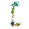
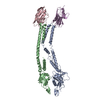
Entry Database : PDB / ID : 7s7kTitle Crystal structure of the EphB2 extracellular domain Ephrin type-B receptor 2 Keywords / / / Function / homology Function Domain/homology Component
/ / / / / / / / / / / / / / / / / / / / / / / / / / / / / / / / / / / / / / / / / / / / / / / / / / / / / / / / / / / / / / / / / / / / / / / / / / / / / / / / / / / / / / / / / / / / / / / / / / / / / / / / / / / / / / / / / / / / / / / / / / / / / / / / / / / / / / / / / / / / / / / / / / / / / / Biological species Mus musculus (house mouse)Method / / / Resolution : 3.15 Å Authors Xu, Y. / Xu, K. / Nikolov, D.B. Funding support Organization Grant number Country National Institutes of Health/National Cancer Institute (NIH/NCI)
Journal : Int J Mol Sci / Year : 2021Title : The Ephb2 Receptor Uses Homotypic, Head-to-Tail Interactions within Its Ectodomain as an Autoinhibitory Control Mechanism.Authors : Xu, Y. / Robev, D. / Saha, N. / Wang, B. / Dalva, M.B. / Xu, K. / Himanen, J.P. / Nikolov, D.B. History Deposition Sep 16, 2021 Deposition site / Processing site Revision 1.0 Oct 27, 2021 Provider / Type Revision 1.1 Oct 18, 2023 Group / Refinement descriptionCategory / chem_comp_bond / pdbx_initial_refinement_modelRevision 1.2 Nov 13, 2024 Group / Category / pdbx_modification_feature / Item
Show all Show less
 Movie
Movie Controller


 Open data
 Basic information
 Components
 Keywords
 Function and homology information
 X-RAY DIFFRACTION /
  Authors
 United States, 1items
  Citation
 Structure visualization
 Downloads & links
 Other downloads


 PDBj
PDBj


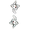
 Assembly
 Trichoplusia ni (cabbage looper)
 Type: D-saccharide, beta linking / Mass: 221.208 Da / Num. of mol.: 2 / Source method: obtained synthetically / Formula: C8H15NO6
 Sample preparation
 Processing