 Movie
Movie Controller

[English] 日本語
 Yorodumi
- PDB-7s6f: Crystal structure of UrtA1 from Synechococcus WH8102 in complex w... -
+ Open data

- Basic information
| Entry | Database: PDB / ID: 7s6f | |||||||||
|---|---|---|---|---|---|---|---|---|---|---|
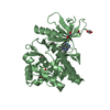
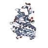
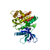
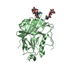
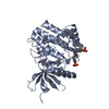
| Title | Crystal structure of UrtA1 from Synechococcus WH8102 in complex with urea and calcium | |||||||||
 Components Components | Putative urea ABC transporter, urea binding protein | |||||||||
 Keywords Keywords | TRANSPORT PROTEIN / Substrate-binding protein / marine cyanobacteria / urea-binding protein | |||||||||
| Function / homology |  Function and homology information Function and homology information | |||||||||
| Biological species |  Synechococcus sp. (bacteria) Synechococcus sp. (bacteria) | |||||||||
| Method |  X-RAY DIFFRACTION / SYNCHROTRON / SAD / Resolution: 1.8 Å X-RAY DIFFRACTION / SYNCHROTRON / SAD / Resolution: 1.8 Å | |||||||||
 Authors Authors | Shah, B.S. / Mikolajek, H. / Orr, C.M. / Mykhaylyk, V. / Owens, R.J. / Paulsen, I.T. | |||||||||
| Funding support |  Australia, 2items Australia, 2items
| |||||||||
 Citation Citation | Journal: To Be Published Title: Crystal structure of UrtA1 from Synechococcus WH8102 in complex with urea and calcium Authors: Shah, B.S. / Mikolajek, H. / Orr, C.M. / Mykhaylyk, V. / Owens, R. / Paulsen, I.T. | |||||||||
| History |
|
- Structure visualization
| Structure viewer | Molecule: MolmilJmol/JSmol |
|---|
- Downloads & links
-Download
| PDBx/mmCIF format | 7s6f.cif.gz | 168.5 KB | Display | PDBx/mmCIF format |
|---|---|---|---|---|
| PDB format | pdb7s6f.ent.gz | 128 KB | Display | PDB format |
| PDBx/mmJSON format | 7s6f.json.gz | Tree view | PDBx/mmJSON format | |
| Others |  Other downloads Other downloads |
-Validation report
| Arichive directory | https://data.pdbj.org/pub/pdb/validation_reports/s6/7s6fftp://data.pdbj.org/pub/pdb/validation_reports/s6/7s6f | HTTPS FTP |
|---|
-Related structure data
| Similar structure data |
|---|


-Links
 PDBj
PDBj


- Assembly
| Deposited unit | 
| ||||||||
|---|---|---|---|---|---|---|---|---|---|
| 1 |
| ||||||||
| Unit cell |
| ||||||||
| Components on special symmetry positions |
|
-Components
| #1: Protein | Mass: 44469.477 Da / Num. of mol.: 1 Source method: isolated from a genetically manipulated source Source: (gene. exp.) Synechococcus sp. (strain WH8102) (bacteria)Strain: WH8102 / Gene: urtA1, SYNW2442 / Plasmid: pOPINM / Details (production host): Addgene Plasmid #26044 / Production host: | ||||||||
|---|---|---|---|---|---|---|---|---|---|
| #2: Chemical | ChemComp-URE /   Mass: 60.055 Da / Num. of mol.: 1 / Source method: obtained synthetically / Formula: CH4N2O / Feature type: SUBJECT OF INVESTIGATION Mass: 60.055 Da / Num. of mol.: 1 / Source method: obtained synthetically / Formula: CH4N2O / Feature type: SUBJECT OF INVESTIGATION | ||||||||
| #3: Chemical |   Mass: 62.068 Da / Num. of mol.: 2 / Source method: obtained synthetically / Formula: C2H6O2 Mass: 62.068 Da / Num. of mol.: 2 / Source method: obtained synthetically / Formula: C2H6O2#4: Chemical | ChemComp-CA /   Mass: 40.078 Da / Num. of mol.: 5 / Source method: obtained synthetically / Formula: Ca / Feature type: SUBJECT OF INVESTIGATION Mass: 40.078 Da / Num. of mol.: 5 / Source method: obtained synthetically / Formula: Ca / Feature type: SUBJECT OF INVESTIGATION#5: Water | ChemComp-HOH / |  Mass: 18.015 Da / Num. of mol.: 263 / Source method: isolated from a natural source / Formula: H2O Mass: 18.015 Da / Num. of mol.: 263 / Source method: isolated from a natural source / Formula: H2OHas ligand of interest | Y | Has protein modification | Y | |
-Experimental details
-Experiment
| Experiment | Method: X-RAY DIFFRACTION / Number of used crystals: 1 |
|---|
- Sample preparation
| Crystal | Density Matthews: 2.29 Å3/Da / Density % sol: 46.53 % |
|---|---|
| Crystal grow | Temperature: 295 K / Method: vapor diffusion, sitting drop / pH: 7.5 Details: 0.05 M Sodium Hepes: MOPS pH 7.5, 0.03 MgCl2, 0.03 CaCl2; 20 % PEG 500 MME; 10 % Peg 20000 Cryo: 30 % Ethylene Glycol |
-Data collection
| Diffraction | Mean temperature: 50 K / Serial crystal experiment: N | ||||||||||||||||||||||||
|---|---|---|---|---|---|---|---|---|---|---|---|---|---|---|---|---|---|---|---|---|---|---|---|---|---|
| Diffraction source | Source: SYNCHROTRON / Site: Diamond  / Beamline: I23 / Wavelength: 2.7552 Å / Beamline: I23 / Wavelength: 2.7552 Å | ||||||||||||||||||||||||
| Detector | Type: DECTRIS PILATUS 12M / Detector: PIXEL / Date: Feb 27, 2020 | ||||||||||||||||||||||||
| Radiation | Monochromator: Si(111) / Protocol: SINGLE WAVELENGTH / Monochromatic (M) / Laue (L): M / Scattering type: x-ray | ||||||||||||||||||||||||
| Radiation wavelength | Wavelength: 2.7552 Å / Relative weight: 1 | ||||||||||||||||||||||||
| Reflection | Resolution: 1.8→120.46 Å / Num. obs: 35399 / % possible obs: 91.3 % / Redundancy: 38.4 % / CC1/2: 0.999 / Rmerge(I) obs: 0.156 / Rpim(I) all: 0.032 / Rrim(I) all: 0.16 / Net I/σ(I): 18 | ||||||||||||||||||||||||
| Reflection shell | Diffraction-ID: 1
|
- Processing
| Software |
| |||||||||||||||||||||||||||||||||||||||||||||||||||||||||||||||||||||||||||||||||||||||||||||||||||||||||||||||||||||||||||||||||||||||||||||||||||||||||||||||||||||
|---|---|---|---|---|---|---|---|---|---|---|---|---|---|---|---|---|---|---|---|---|---|---|---|---|---|---|---|---|---|---|---|---|---|---|---|---|---|---|---|---|---|---|---|---|---|---|---|---|---|---|---|---|---|---|---|---|---|---|---|---|---|---|---|---|---|---|---|---|---|---|---|---|---|---|---|---|---|---|---|---|---|---|---|---|---|---|---|---|---|---|---|---|---|---|---|---|---|---|---|---|---|---|---|---|---|---|---|---|---|---|---|---|---|---|---|---|---|---|---|---|---|---|---|---|---|---|---|---|---|---|---|---|---|---|---|---|---|---|---|---|---|---|---|---|---|---|---|---|---|---|---|---|---|---|---|---|---|---|---|---|---|---|---|---|---|---|
| Refinement | Method to determine structure: SAD / Resolution: 1.8→71.492 Å / Cor.coef. Fo:Fc: 0.974 / Cor.coef. Fo:Fc free: 0.958 / SU B: 2.849 / SU ML: 0.084 / Cross valid method: FREE R-VALUE / ESU R: 0.122 / ESU R Free: 0.113 Details: Hydrogens have been added in their riding positions
| |||||||||||||||||||||||||||||||||||||||||||||||||||||||||||||||||||||||||||||||||||||||||||||||||||||||||||||||||||||||||||||||||||||||||||||||||||||||||||||||||||||
| Solvent computation | Ion probe radii: 0.8 Å / Shrinkage radii: 0.8 Å / VDW probe radii: 1.2 Å / Solvent model: MASK BULK SOLVENT | |||||||||||||||||||||||||||||||||||||||||||||||||||||||||||||||||||||||||||||||||||||||||||||||||||||||||||||||||||||||||||||||||||||||||||||||||||||||||||||||||||||
| Displacement parameters | Biso mean: 33.343 Å2
| |||||||||||||||||||||||||||||||||||||||||||||||||||||||||||||||||||||||||||||||||||||||||||||||||||||||||||||||||||||||||||||||||||||||||||||||||||||||||||||||||||||
| Refinement step | Cycle: LAST / Resolution: 1.8→71.492 Å
| |||||||||||||||||||||||||||||||||||||||||||||||||||||||||||||||||||||||||||||||||||||||||||||||||||||||||||||||||||||||||||||||||||||||||||||||||||||||||||||||||||||
| Refine LS restraints |
| |||||||||||||||||||||||||||||||||||||||||||||||||||||||||||||||||||||||||||||||||||||||||||||||||||||||||||||||||||||||||||||||||||||||||||||||||||||||||||||||||||||
| LS refinement shell |
|
