 Movie
Movie Controller

[English] 日本語
 Yorodumi
- PDB-7s69: N-acetylglucosamine-1-phosphotransferase (GNPT) gamma subunit (GN... -
+ Open data

- Basic information
| Entry | Database: PDB / ID: 7s69 | ||||||
|---|---|---|---|---|---|---|---|
| Title | N-acetylglucosamine-1-phosphotransferase (GNPT) gamma subunit (GNPTG), from clawed frog | ||||||
 Components Components | N-acetylglucosamine-1-phosphotransferase gamma subunit | ||||||
 Keywords Keywords | SUGAR BINDING PROTEIN / GNPT / mannose / mannose 6-phosphate receptor homology (MRH) domain / mucolipidosis | ||||||
| Function / homology | Glucosidase 2 subunit beta-like / Glucosidase II beta subunit-like / Glucosidase II beta subunit-like protein / DMAP1-binding domain / DMAP1-binding domain profile. / Mannose-6-phosphate receptor binding domain superfamily / MRH domain / MRH domain profile. / LOC446283 protein Function and homology information Function and homology information | ||||||
| Biological species | |||||||
| Method |  X-RAY DIFFRACTION / SYNCHROTRON / MOLECULAR REPLACEMENT / Resolution: 3.04 Å X-RAY DIFFRACTION / SYNCHROTRON / MOLECULAR REPLACEMENT / Resolution: 3.04 Å | ||||||
 Authors Authors | Gorelik, A. / Illes, K. / Nagar, B. | ||||||
| Funding support |  Canada, 1items Canada, 1items
| ||||||
 Citation Citation | Journal: Proc.Natl.Acad.Sci.USA / Year: 2022 Title: Structures of the mannose-6-phosphate pathway enzyme, GlcNAc-1-phosphotransferase. Authors: Gorelik, A. / Illes, K. / Bui, K.H. / Nagar, B. | ||||||
| History |
|
- Structure visualization
| Structure viewer | Molecule: MolmilJmol/JSmol |
|---|
- Downloads & links
-Download
| PDBx/mmCIF format | 7s69.cif.gz | 158.1 KB | Display | PDBx/mmCIF format |
|---|---|---|---|---|
| PDB format | pdb7s69.ent.gz | 123.3 KB | Display | PDB format |
| PDBx/mmJSON format | 7s69.json.gz | Tree view | PDBx/mmJSON format | |
| Others |  Other downloads Other downloads |
-Validation report
| Arichive directory | https://data.pdbj.org/pub/pdb/validation_reports/s6/7s69ftp://data.pdbj.org/pub/pdb/validation_reports/s6/7s69 | HTTPS FTP |
|---|
-Related structure data
| Related structure data | 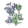 7s6nC 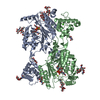 7sj2C  2lvxS S: Starting model for refinement C: citing same article ( |
|---|---|
| Similar structure data |
-Links
 PDBj
PDBj
- Assembly
| Deposited unit | 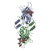
| ||||||||||||
|---|---|---|---|---|---|---|---|---|---|---|---|---|---|
| 1 |
| ||||||||||||
| Unit cell |
|
-Components
| #1: Protein | Mass: 33412.453 Da / Num. of mol.: 2 Source method: isolated from a genetically manipulated source Source: (gene. exp.)   Spodoptera frugiperda (fall armyworm) / References: UniProt: Q68F17 Spodoptera frugiperda (fall armyworm) / References: UniProt: Q68F17#2: Polysaccharide | alpha-D-mannopyranose-(1-3)-alpha-D-mannopyranose-(1-6)-[alpha-D-mannopyranose-(1-3)]beta-D- ...alpha-D-mannopyranose-(1-3)-alpha-D-mannopyranose-(1-6)-[alpha-D-mannopyranose-(1-3)]beta-D-mannopyranose-(1-4)-2-acetamido-2-deoxy-beta-D-glucopyranose-(1-4)-2-acetamido-2-deoxy-beta-D-glucopyranose | Source method: isolated from a genetically manipulated source #3: Polysaccharide | alpha-D-mannopyranose-(1-2)-alpha-D-mannopyranose-(1-6)-[alpha-D-mannopyranose-(1-3)]alpha-D- ...alpha-D-mannopyranose-(1-2)-alpha-D-mannopyranose-(1-6)-[alpha-D-mannopyranose-(1-3)]alpha-D-mannopyranose-(1-6)-[alpha-D-mannopyranose-(1-2)-alpha-D-mannopyranose-(1-3)]beta-D-mannopyranose-(1-4)-2-acetamido-2-deoxy-beta-D-glucopyranose-(1-4)-2-acetamido-2-deoxy-beta-D-glucopyranose | Source method: isolated from a genetically manipulated source #4: Chemical | ChemComp-CL /   Mass: 35.453 Da / Num. of mol.: 10 / Source method: obtained synthetically / Formula: Cl Mass: 35.453 Da / Num. of mol.: 10 / Source method: obtained synthetically / Formula: Cl#5: Water | ChemComp-HOH / |  Mass: 18.015 Da / Num. of mol.: 29 / Source method: isolated from a natural source / Formula: H2O Mass: 18.015 Da / Num. of mol.: 29 / Source method: isolated from a natural source / Formula: H2OHas ligand of interest | Y | Has protein modification | Y | |
|---|
-Experimental details
-Experiment
| Experiment | Method: X-RAY DIFFRACTION / Number of used crystals: 1 |
|---|
- Sample preparation
| Crystal | Density Matthews: 1.89 Å3/Da / Density % sol: 35.02 % |
|---|---|
| Crystal grow | Temperature: 293 K / Method: vapor diffusion, sitting drop / pH: 8 / Details: 20% PEG 3350, 0.1M bis-tris propane pH8, 0.2M KNO3 |
-Data collection
| Diffraction | Mean temperature: 100 K / Serial crystal experiment: N |
|---|---|
| Diffraction source | Source: SYNCHROTRON / Site: ALS  / Beamline: 5.0.2 / Wavelength: 1 Å / Beamline: 5.0.2 / Wavelength: 1 Å |
| Detector | Type: DECTRIS PILATUS3 S 6M / Detector: PIXEL / Date: Dec 3, 2018 |
| Radiation | Protocol: SINGLE WAVELENGTH / Monochromatic (M) / Laue (L): M / Scattering type: x-ray |
| Radiation wavelength | Wavelength: 1 Å / Relative weight: 1 |
| Reflection | Resolution: 3.04→50 Å / Num. obs: 8751 / % possible obs: 85 % / Redundancy: 8 % / Biso Wilson estimate: 58.1 Å2 / Rrim(I) all: 0.136 / Net I/σ(I): 12.7 |
| Reflection shell | Resolution: 3.04→3.11 Å / Mean I/σ(I) obs: 1.29 / Num. unique obs: 577 / CC1/2: 0.812 |
- Processing
| Software |
| |||||||||||||||||||||||||||||||||||||||||||||||||
|---|---|---|---|---|---|---|---|---|---|---|---|---|---|---|---|---|---|---|---|---|---|---|---|---|---|---|---|---|---|---|---|---|---|---|---|---|---|---|---|---|---|---|---|---|---|---|---|---|---|---|
| Refinement | Method to determine structure: MOLECULAR REPLACEMENT Starting model: 2LVX Resolution: 3.04→49.19 Å / SU ML: 0.3467 / Cross valid method: FREE R-VALUE / σ(F): 1.35 / Phase error: 24.0518 Stereochemistry target values: GeoStd + Monomer Library + CDL v1.2
| |||||||||||||||||||||||||||||||||||||||||||||||||
| Solvent computation | Shrinkage radii: 0.9 Å / VDW probe radii: 1.11 Å / Solvent model: FLAT BULK SOLVENT MODEL | |||||||||||||||||||||||||||||||||||||||||||||||||
| Displacement parameters | Biso mean: 56.35 Å2 | |||||||||||||||||||||||||||||||||||||||||||||||||
| Refinement step | Cycle: LAST / Resolution: 3.04→49.19 Å
| |||||||||||||||||||||||||||||||||||||||||||||||||
| Refine LS restraints |
| |||||||||||||||||||||||||||||||||||||||||||||||||
| LS refinement shell |
|
