 Movie
Movie Controller

[English] 日本語
 Yorodumi
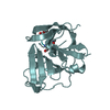
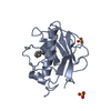
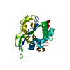
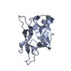
- PDB-7s54: Sortase A from Streptococcus agalactiae with the deltaN188 b7-b8 ... -
+ Open data

- Basic information
| Entry | Database: PDB / ID: 7s54 | ||||||
|---|---|---|---|---|---|---|---|
| Title | Sortase A from Streptococcus agalactiae with the deltaN188 b7-b8 loop sequence from Staphylococcus aureus Sortase A | ||||||
 Components Components | Class A sortase, sortase A chimera | ||||||
 Keywords Keywords | HYDROLASE / sortase-fold / sortase / eight-stranded beta barrel / transpeptidase / housekeeping sortase / surface protein | ||||||
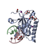
| Function / homology |  Function and homology information Function and homology informationsortase A / cysteine-type peptidase activity / proteolysis / metal ion binding / plasma membrane Similarity search - Function | ||||||
| Biological species |  Streptococcus agalactiae (bacteria) Streptococcus agalactiae (bacteria) Staphylococcus aureus (bacteria) Staphylococcus aureus (bacteria) | ||||||
| Method |  X-RAY DIFFRACTION / SYNCHROTRON / MOLECULAR REPLACEMENT / Resolution: 1.794 Å X-RAY DIFFRACTION / SYNCHROTRON / MOLECULAR REPLACEMENT / Resolution: 1.794 Å | ||||||
 Authors Authors | Gao, M. / Kodama, H.M. / Antos, J.M. / Amacher, J.F. | ||||||
| Funding support |  United States, 1items United States, 1items
| ||||||
 Citation Citation | Journal: Protein Sci. / Year: 2022 Title: Structural and biochemical analyses of selectivity determinants in chimeric Streptococcus Class A sortase enzymes. Authors: Gao, M. / Johnson, D.A. / Piper, I.M. / Kodama, H.M. / Svendsen, J.E. / Tahti, E. / Longshore-Neate, F. / Vogel, B. / Antos, J.M. / Amacher, J.F. | ||||||
| History |
|
- Structure visualization
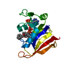
| Structure viewer | Molecule: MolmilJmol/JSmol |
|---|
- Downloads & links
-Download
| PDBx/mmCIF format | 7s54.cif.gz | 83.5 KB | Display | PDBx/mmCIF format |
|---|---|---|---|---|
| PDB format | pdb7s54.ent.gz | 61 KB | Display | PDB format |
| PDBx/mmJSON format | 7s54.json.gz | Tree view | PDBx/mmJSON format | |
| Others |  Other downloads Other downloads |
-Validation report
| Arichive directory | https://data.pdbj.org/pub/pdb/validation_reports/s5/7s54ftp://data.pdbj.org/pub/pdb/validation_reports/s5/7s54 | HTTPS FTP |
|---|
-Related structure data
| Related structure data | 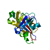 7s53C 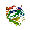 7s56C 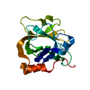 7s57C  3rccS S: Starting model for refinement C: citing same article ( |
|---|---|
| Similar structure data |


-Links
 PDBj
PDBj- Assembly
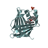
| Deposited unit | 
| ||||||||
|---|---|---|---|---|---|---|---|---|---|
| 1 | 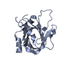
| ||||||||
| 2 | 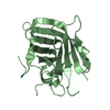
| ||||||||
| Unit cell |
|
-Components
| #1: Protein | Mass: 19027.703 Da / Num. of mol.: 2 Source method: isolated from a genetically manipulated source Source: (gene. exp.) Streptococcus agalactiae (bacteria), (gene. exp.) Staphylococcus aureus (strain NCTC 8325 / PS 47) (bacteria)Gene: srtA, C4618_06055, F5043_06280, GD434_06060, NCTC6175_01342, RDF_0944, srtA, SAOUHSC_02834 Plasmid: pET28a(+) / Strain: NCTC 8325 / PS 47 / Production host: #2: Water | ChemComp-HOH / |  Mass: 18.015 Da / Num. of mol.: 213 / Source method: isolated from a natural source / Formula: H2O Mass: 18.015 Da / Num. of mol.: 213 / Source method: isolated from a natural source / Formula: H2O |
|---|
-Experimental details
-Experiment
| Experiment | Method: X-RAY DIFFRACTION / Number of used crystals: 1 |
|---|
- Sample preparation
| Crystal | Density Matthews: 1.81 Å3/Da / Density % sol: 32.23 % |
|---|---|
| Crystal grow | Temperature: 298 K / Method: vapor diffusion, hanging drop / pH: 6 Details: 0.02 MES monohydrate pH 6, 12% (v/v) 2-Propanol, 24% (w/v) PEG monomethyl ether 2000 |
-Data collection
| Diffraction | Mean temperature: 80 K / Serial crystal experiment: N | ||||||||||||||||||||||||
|---|---|---|---|---|---|---|---|---|---|---|---|---|---|---|---|---|---|---|---|---|---|---|---|---|---|
| Diffraction source | Source: SYNCHROTRON / Site: ALS / Beamline: 5.0.1 / Wavelength: 0.97741 Å | ||||||||||||||||||||||||
| Detector | Type: DECTRIS PILATUS3 6M / Detector: PIXEL / Date: Apr 14, 2021 | ||||||||||||||||||||||||
| Radiation | Protocol: SINGLE WAVELENGTH / Monochromatic (M) / Laue (L): M / Scattering type: x-ray | ||||||||||||||||||||||||
| Radiation wavelength | Wavelength: 0.97741 Å / Relative weight: 1 | ||||||||||||||||||||||||
| Reflection | Resolution: 1.79→35.631 Å / Num. obs: 25832 / % possible obs: 99 % / Redundancy: 6.6 % / Biso Wilson estimate: 17.89 Å2 / CC1/2: 0.996 / Rmerge(I) obs: 0.148 / Rpim(I) all: 0.062 / Rrim(I) all: 0.16 / Net I/σ(I): 8.4 | ||||||||||||||||||||||||
| Reflection shell | Diffraction-ID: 1
|
- Processing
| Software |
| ||||||||||||||||||||||||||||||||||||||||||||||||||||||||||||
|---|---|---|---|---|---|---|---|---|---|---|---|---|---|---|---|---|---|---|---|---|---|---|---|---|---|---|---|---|---|---|---|---|---|---|---|---|---|---|---|---|---|---|---|---|---|---|---|---|---|---|---|---|---|---|---|---|---|---|---|---|---|
| Refinement | Method to determine structure: MOLECULAR REPLACEMENT Starting model: 3RCC Resolution: 1.794→35.631 Å / SU ML: 0.24 / Cross valid method: THROUGHOUT / σ(F): 1.34 / Phase error: 27.82 / Stereochemistry target values: ML
| ||||||||||||||||||||||||||||||||||||||||||||||||||||||||||||
| Solvent computation | Shrinkage radii: 0.9 Å / VDW probe radii: 1.11 Å / Solvent model: FLAT BULK SOLVENT MODEL | ||||||||||||||||||||||||||||||||||||||||||||||||||||||||||||
| Displacement parameters | Biso max: 67.6 Å2 / Biso mean: 25.8436 Å2 / Biso min: 8.58 Å2 | ||||||||||||||||||||||||||||||||||||||||||||||||||||||||||||
| Refinement step | Cycle: final / Resolution: 1.794→35.631 Å
| ||||||||||||||||||||||||||||||||||||||||||||||||||||||||||||
| Refine LS restraints |
| ||||||||||||||||||||||||||||||||||||||||||||||||||||||||||||
| LS refinement shell | Refine-ID: X-RAY DIFFRACTION / Rfactor Rfree error: 0
|
