 Movie
Movie Controller

[English] 日本語
 Yorodumi
- PDB-7s0b: Structure of the SARS-CoV-2 RBD in complex with neutralizing anti... -
+ Open data

- Basic information
| Entry | Database: PDB / ID: 7s0b | ||||||
|---|---|---|---|---|---|---|---|
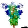
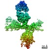
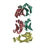
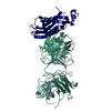
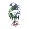
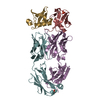
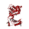
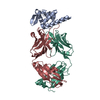
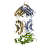
| Title | Structure of the SARS-CoV-2 RBD in complex with neutralizing antibody N-612-056 | ||||||
 Components Components |
| ||||||
 Keywords Keywords | VIRAL PROTEIN/IMMUNE SYSTEM / Antibody / SARS-CoV-2 / COVID-19 / mRNA Display / ANTIVIRAL PROTEIN / VIRAL PROTEIN-IMMUNE SYSTEM complex | ||||||
| Function / homology |  Function and homology information Function and homology informationsymbiont-mediated disruption of host tissue / Maturation of spike protein / Translation of Structural Proteins / Virion Assembly and Release / host cell surface / host extracellular space / viral translation / symbiont-mediated-mediated suppression of host tetherin activity / Induction of Cell-Cell Fusion / structural constituent of virion ...symbiont-mediated disruption of host tissue / Maturation of spike protein / Translation of Structural Proteins / Virion Assembly and Release / host cell surface / host extracellular space / viral translation / symbiont-mediated-mediated suppression of host tetherin activity / Induction of Cell-Cell Fusion / structural constituent of virion / entry receptor-mediated virion attachment to host cell / membrane fusion / Attachment and Entry / host cell endoplasmic reticulum-Golgi intermediate compartment membrane / positive regulation of viral entry into host cell / receptor-mediated virion attachment to host cell / host cell surface receptor binding / symbiont-mediated suppression of host innate immune response / receptor ligand activity / endocytosis involved in viral entry into host cell / fusion of virus membrane with host plasma membrane / fusion of virus membrane with host endosome membrane / viral envelope / symbiont entry into host cell / virion attachment to host cell / SARS-CoV-2 activates/modulates innate and adaptive immune responses / host cell plasma membrane / virion membrane / identical protein binding / membrane / plasma membrane Similarity search - Function | ||||||
| Biological species |  Homo sapiens (human) Homo sapiens (human)  Severe acute respiratory syndrome coronavirus 2 Severe acute respiratory syndrome coronavirus 2 | ||||||
| Method |  X-RAY DIFFRACTION / SYNCHROTRON / MOLECULAR REPLACEMENT / Resolution: 2.9 Å X-RAY DIFFRACTION / SYNCHROTRON / MOLECULAR REPLACEMENT / Resolution: 2.9 Å | ||||||
 Authors Authors | Tanaka, S. / Barnes, C.O. / Bjorkman, P.J. | ||||||
| Funding support |  United States, 1items United States, 1items
| ||||||
 Citation Citation | Journal: Cell Rep / Year: 2022 Title: Rapid identification of neutralizing antibodies against SARS-CoV-2 variants by mRNA display. Authors: Shiho Tanaka / C Anders Olson / Christopher O Barnes / Wendy Higashide / Marcos Gonzalez / Justin Taft / Ashley Richardson / Marta Martin-Fernandez / Dusan Bogunovic / Priyanthi N P ...Authors: Shiho Tanaka / C Anders Olson / Christopher O Barnes / Wendy Higashide / Marcos Gonzalez / Justin Taft / Ashley Richardson / Marta Martin-Fernandez / Dusan Bogunovic / Priyanthi N P Gnanapragasam / Pamela J Bjorkman / Patricia Spilman / Kayvan Niazi / Shahrooz Rabizadeh / Patrick Soon-Shiong / Abstract: The increasing prevalence of severe acute respiratory syndrome coronavirus 2 (SARS-CoV-2) variants with the ability to escape existing humoral protection conferred by previous infection and/or ...The increasing prevalence of severe acute respiratory syndrome coronavirus 2 (SARS-CoV-2) variants with the ability to escape existing humoral protection conferred by previous infection and/or immunization necessitates the discovery of broadly reactive neutralizing antibodies (nAbs). Utilizing mRNA display, we identify a set of antibodies against SARS-CoV-2 spike (S) proteins and characterize the structures of nAbs that recognize epitopes in the S1 subunit of the S glycoprotein. These structural studies reveal distinct binding modes for several antibodies, including the targeting of rare cryptic epitopes in the receptor-binding domain (RBD) of S that interact with angiotensin-converting enzyme 2 (ACE2) to initiate infection, as well as the S1 subdomain 1. Further, we engineer a potent ACE2-blocking nAb to sustain binding to S RBD with the E484K and L452R substitutions found in multiple SARS-CoV-2 variants. We demonstrate that mRNA display is an approach for the rapid identification of nAbs that can be used in combination to combat emerging SARS-CoV-2 variants. #1: Journal: bioRxiv / Year: 2021 Title: Rapid Identification of Neutralizing Antibodies against SARS-CoV-2 Variants by mRNA Display. Authors: Shiho Tanaka / C Anders Olson / Christopher O Barnes / Wendy Higashide / Marcos Gonzalez / Justin Taft / Ashley Richardson / Marta Martin-Fernandez / Dusan Bogunovic / Priyanthi N P ...Authors: Shiho Tanaka / C Anders Olson / Christopher O Barnes / Wendy Higashide / Marcos Gonzalez / Justin Taft / Ashley Richardson / Marta Martin-Fernandez / Dusan Bogunovic / Priyanthi N P Gnanapragasam / Pamela J Bjorkman / Patricia Spilman / Kayvan Niazi / Shahrooz Rabizadeh / Patrick Soon-Shiong Abstract: The increasing prevalence of SARS-CoV-2 variants with the ability to escape existing humoral protection conferred by previous infection and/or immunization necessitates the discovery of broadly- ...The increasing prevalence of SARS-CoV-2 variants with the ability to escape existing humoral protection conferred by previous infection and/or immunization necessitates the discovery of broadly-reactive neutralizing antibodies (nAbs). Utilizing mRNA display, we identified a set of antibodies against SARS-CoV-2 spike (S) proteins and characterized the structures of nAbs that recognized epitopes in the S1 subunit of the S glycoprotein. These structural studies revealed distinct binding modes for several antibodies, including targeting of rare cryptic epitopes in the receptor-binding domain (RBD) of S that interacts with angiotensin- converting enzyme 2 (ACE2) to initiate infection, as well as the S1 subdomain 1. A potent ACE2-blocking nAb was further engineered to sustain binding to S RBD with the E484K and L452R substitutions found in multiple SARS-CoV-2 variants. We demonstrate that mRNA display is a promising approach for the rapid identification of nAbs that can be used in combination to combat emerging SARS-CoV-2 variants. | ||||||
| History |
|
- Structure visualization
| Structure viewer | Molecule: MolmilJmol/JSmol |
|---|
- Downloads & links
-Download
| PDBx/mmCIF format | 7s0b.cif.gz | 594.9 KB | Display | PDBx/mmCIF format |
|---|---|---|---|---|
| PDB format | pdb7s0b.ent.gz | 411.3 KB | Display | PDB format |
| PDBx/mmJSON format | 7s0b.json.gz | Tree view | PDBx/mmJSON format | |
| Others |  Other downloads Other downloads |
-Validation report
| Summary document | 7s0b_validation.pdf.gz | 505.2 KB | Display | wwPDB validaton report |
|---|---|---|---|---|
| Full document | 7s0b_full_validation.pdf.gz | 531.3 KB | Display | |
| Data in XML | 7s0b_validation.xml.gz | 47.5 KB | Display | |
| Data in CIF | 7s0b_validation.cif.gz | 65.2 KB | Display | |
| Arichive directory | https://data.pdbj.org/pub/pdb/validation_reports/s0/7s0bftp://data.pdbj.org/pub/pdb/validation_reports/s0/7s0b | HTTPS FTP |
-Related structure data
| Related structure data |  7s0cC  7s0dC  7s0eC 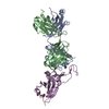 7k8mS S: Starting model for refinement C: citing same article ( |
|---|---|
| Similar structure data |


-Links
 PDBj
PDBj


- Assembly
| Deposited unit | 
| |||||||||||||||||||||||||||||||||||||||||||||||||||||||||||||||||||||||||||||||||||||||||||||||||||||||||||||||||||||||||||
|---|---|---|---|---|---|---|---|---|---|---|---|---|---|---|---|---|---|---|---|---|---|---|---|---|---|---|---|---|---|---|---|---|---|---|---|---|---|---|---|---|---|---|---|---|---|---|---|---|---|---|---|---|---|---|---|---|---|---|---|---|---|---|---|---|---|---|---|---|---|---|---|---|---|---|---|---|---|---|---|---|---|---|---|---|---|---|---|---|---|---|---|---|---|---|---|---|---|---|---|---|---|---|---|---|---|---|---|---|---|---|---|---|---|---|---|---|---|---|---|---|---|---|---|---|
| 1 | 
| |||||||||||||||||||||||||||||||||||||||||||||||||||||||||||||||||||||||||||||||||||||||||||||||||||||||||||||||||||||||||||
| 2 | 
| |||||||||||||||||||||||||||||||||||||||||||||||||||||||||||||||||||||||||||||||||||||||||||||||||||||||||||||||||||||||||||
| Unit cell |
| |||||||||||||||||||||||||||||||||||||||||||||||||||||||||||||||||||||||||||||||||||||||||||||||||||||||||||||||||||||||||||
| Noncrystallographic symmetry (NCS) | NCS domain:
NCS domain segments:
|