 Movie
Movie Controller

[English] 日本語
 Yorodumi
- PDB-7s04: 1-deoxy-D-xylulose 5-phosphate reductoisomerase (IspC) from Acine... -
+ Open data

- Basic information
| Entry | Database: PDB / ID: 7s04 | |||||||||
|---|---|---|---|---|---|---|---|---|---|---|
| Title | 1-deoxy-D-xylulose 5-phosphate reductoisomerase (IspC) from Acinetobacter baumannii in complex with FR900098, NADPH, and a magnesium ion | |||||||||
 Components Components | 1-deoxy-D-xylulose 5-phosphate reductoisomerase | |||||||||
 Keywords Keywords | OXIDOREDUCTASE/INHIBITOR / isoprenoid biosynthesis / inhibitor / complex / OXIDOREDUCTASE-INHIBITOR complex | |||||||||
| Function / homology |  Function and homology information Function and homology information: / 1-deoxy-D-xylulose-5-phosphate reductoisomerase / 1-deoxy-D-xylulose-5-phosphate reductoisomerase activity / isomerase activity / NADPH binding / manganese ion binding Similarity search - Function | |||||||||
| Biological species |  Acinetobacter baumannii (bacteria) Acinetobacter baumannii (bacteria) | |||||||||
| Method |  X-RAY DIFFRACTION / MOLECULAR REPLACEMENT / Resolution: 2.52 Å X-RAY DIFFRACTION / MOLECULAR REPLACEMENT / Resolution: 2.52 Å | |||||||||
 Authors Authors | Noble, S.M. / Ball, H.S. / Couch, R.D. | |||||||||
| Funding support | 2items
| |||||||||
 Citation Citation | Journal: Acs Infect Dis. / Year: 2021 Title: Characterization and Inhibition of 1-Deoxy-d-Xylulose 5-Phosphate Reductoisomerase: A Promising Drug Target in Acinetobacter baumannii and Klebsiella pneumoniae . Authors: Ball, H.S. / Girma, M.B. / Zainab, M. / Soojhawon, I. / Couch, R.D. / Noble, S.M. | |||||||||
| History |
|
- Structure visualization
| Structure viewer | Molecule: MolmilJmol/JSmol |
|---|
- Downloads & links
-Download
| PDBx/mmCIF format | 7s04.cif.gz | 175.7 KB | Display | PDBx/mmCIF format |
|---|---|---|---|---|
| PDB format | pdb7s04.ent.gz | 130.4 KB | Display | PDB format |
| PDBx/mmJSON format | 7s04.json.gz | Tree view | PDBx/mmJSON format | |
| Others |  Other downloads Other downloads |
-Validation report
| Arichive directory | https://data.pdbj.org/pub/pdb/validation_reports/s0/7s04ftp://data.pdbj.org/pub/pdb/validation_reports/s0/7s04 | HTTPS FTP |
|---|
-Related structure data
| Related structure data |  4zn6S S: Starting model for refinement |
|---|---|
| Similar structure data |


-Links
 PDBj
PDBj

- Assembly
| Deposited unit | 
| ||||||||||||
|---|---|---|---|---|---|---|---|---|---|---|---|---|---|
| 1 | 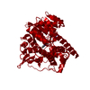
| ||||||||||||
| Unit cell |
| ||||||||||||
| Components on special symmetry positions |
|
-Components
| #1: Protein | Mass: 44095.648 Da / Num. of mol.: 1 Source method: isolated from a genetically manipulated source Source: (gene. exp.) Acinetobacter baumannii (bacteria)Gene: dxr, ABA1_02159, Aba9102_18380, ABUW_1739, AN415_01721, C6N18_11275, DLI69_14610, F2P40_10340, FDO31_03370, GNY86_03270, HYI42_18125, NCTC13421_01630 Production host: References: UniProt: A0A0D5YGN4, 1-deoxy-D-xylulose-5-phosphate reductoisomerase |
|---|---|
| #2: Chemical | ChemComp-F98 /   Mass: 197.126 Da / Num. of mol.: 1 / Source method: obtained synthetically / Formula: C5H12NO5P / Feature type: SUBJECT OF INVESTIGATION Mass: 197.126 Da / Num. of mol.: 1 / Source method: obtained synthetically / Formula: C5H12NO5P / Feature type: SUBJECT OF INVESTIGATION |
| #3: Chemical | ChemComp-MG /   Mass: 24.305 Da / Num. of mol.: 1 / Source method: obtained synthetically / Formula: Mg Mass: 24.305 Da / Num. of mol.: 1 / Source method: obtained synthetically / Formula: Mg |
| #4: Chemical | ChemComp-NDP /   Mass: 745.421 Da / Num. of mol.: 1 / Source method: obtained synthetically / Formula: C21H30N7O17P3 / Feature type: SUBJECT OF INVESTIGATION Mass: 745.421 Da / Num. of mol.: 1 / Source method: obtained synthetically / Formula: C21H30N7O17P3 / Feature type: SUBJECT OF INVESTIGATION |
| #5: Water | ChemComp-HOH /  Mass: 18.015 Da / Num. of mol.: 147 / Source method: isolated from a natural source / Formula: H2O Mass: 18.015 Da / Num. of mol.: 147 / Source method: isolated from a natural source / Formula: H2O |
| Has ligand of interest | Y |
-Experimental details
-Experiment
| Experiment | Method: X-RAY DIFFRACTION / Number of used crystals: 1 |
|---|
- Sample preparation
| Crystal | Density Matthews: 2.41 Å3/Da / Density % sol: 48.88 % |
|---|---|
| Crystal grow | Temperature: 289.15 K / Method: vapor diffusion, sitting drop / pH: 5.6 Details: 100 mM sodium citrate:HCl, pH 5.60, 250 mM ammonium sulfate, 24% PEG4000 |
-Data collection
| Diffraction | Mean temperature: 100 K / Serial crystal experiment: N |
|---|---|
| Diffraction source | Source: ROTATING ANODE / Type: BRUKER AXS MICROSTAR / Wavelength: 1.54 Å |
| Detector | Type: Bruker Platinum 135 / Detector: CCD / Date: Mar 15, 2019 |
| Radiation | Monochromator: M / Protocol: SINGLE WAVELENGTH / Monochromatic (M) / Laue (L): M / Scattering type: x-ray |
| Radiation wavelength | Wavelength: 1.54 Å / Relative weight: 1 |
| Reflection | Resolution: 2.52→33.92 Å / Num. obs: 27390 / % possible obs: 98.61 % / Redundancy: 5.49 % / Biso Wilson estimate: 23.31 Å2 / Rmerge(I) obs: 0.2494 / Net I/σ(I): 23.09 |
| Reflection shell | Resolution: 2.52→2.56 Å / Redundancy: 11.06 % / Rmerge(I) obs: 0.36 / Num. unique obs: 14804 / Rsym value: 0.12 / % possible all: 98 |
- Processing
| Software |
| |||||||||||||||||||||||||||||||||||||||||||||||||||||||||||||||||||||||||||||||||||||||||||||||||||||||||||||||||||||||||||||||||||||||||||||||||||
|---|---|---|---|---|---|---|---|---|---|---|---|---|---|---|---|---|---|---|---|---|---|---|---|---|---|---|---|---|---|---|---|---|---|---|---|---|---|---|---|---|---|---|---|---|---|---|---|---|---|---|---|---|---|---|---|---|---|---|---|---|---|---|---|---|---|---|---|---|---|---|---|---|---|---|---|---|---|---|---|---|---|---|---|---|---|---|---|---|---|---|---|---|---|---|---|---|---|---|---|---|---|---|---|---|---|---|---|---|---|---|---|---|---|---|---|---|---|---|---|---|---|---|---|---|---|---|---|---|---|---|---|---|---|---|---|---|---|---|---|---|---|---|---|---|---|---|---|---|
| Refinement | Method to determine structure: MOLECULAR REPLACEMENT Starting model: PDB entry 4ZN6 Resolution: 2.52→33.92 Å / SU ML: 0.301 / Cross valid method: THROUGHOUT / σ(F): 1.34 / Phase error: 25.7134 / Stereochemistry target values: GeoStd + Monomer Library
| |||||||||||||||||||||||||||||||||||||||||||||||||||||||||||||||||||||||||||||||||||||||||||||||||||||||||||||||||||||||||||||||||||||||||||||||||||
| Solvent computation | Shrinkage radii: 0.9 Å / VDW probe radii: 1.11 Å / Solvent model: FLAT BULK SOLVENT MODEL | |||||||||||||||||||||||||||||||||||||||||||||||||||||||||||||||||||||||||||||||||||||||||||||||||||||||||||||||||||||||||||||||||||||||||||||||||||
| Displacement parameters | Biso mean: 23.65 Å2 | |||||||||||||||||||||||||||||||||||||||||||||||||||||||||||||||||||||||||||||||||||||||||||||||||||||||||||||||||||||||||||||||||||||||||||||||||||
| Refinement step | Cycle: LAST / Resolution: 2.52→33.92 Å
| |||||||||||||||||||||||||||||||||||||||||||||||||||||||||||||||||||||||||||||||||||||||||||||||||||||||||||||||||||||||||||||||||||||||||||||||||||
| Refine LS restraints |
| |||||||||||||||||||||||||||||||||||||||||||||||||||||||||||||||||||||||||||||||||||||||||||||||||||||||||||||||||||||||||||||||||||||||||||||||||||
| LS refinement shell |
|
