 ムービー
ムービー コントローラー


+ データを開く

- 基本情報
基本情報
| 登録情報 | データベース: PDB / ID: 7rwy | ||||||||||||
|---|---|---|---|---|---|---|---|---|---|---|---|---|---|
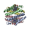
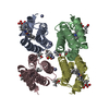
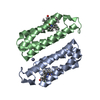
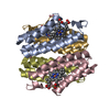
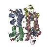
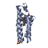
| タイトル | Crystal structure of a Fe-bound RIDC1 variant in the presence of reductant | ||||||||||||
 要素 要素 | Soluble cytochrome b562 | ||||||||||||
 キーワード キーワード | METAL BINDING PROTEIN | ||||||||||||
| 機能・相同性 |  機能・相同性情報 機能・相同性情報electron transport chain / periplasmic space / electron transfer activity / iron ion binding / heme binding 類似検索 - 分子機能 | ||||||||||||
| 生物種 |  | ||||||||||||
| 手法 |  X線回折 / シンクロトロン / 分子置換 / 解像度: 2.2 Å X線回折 / シンクロトロン / 分子置換 / 解像度: 2.2 Å | ||||||||||||
 データ登録者 データ登録者 | Kakkis, A. / Golub, E. | ||||||||||||
| 資金援助 |  米国, 3件 米国, 3件
| ||||||||||||
 引用 引用 | ジャーナル: Chem.Commun.(Camb.) / 年: 2022 タイトル: Redox- and metal-directed structural diversification in designed metalloprotein assemblies. 著者: Kakkis, A. / Golub, E. / Choi, T.S. / Tezcan, F.A. | ||||||||||||
| 履歴 |
|
- 構造の表示
構造の表示
| 構造ビューア | 分子: MolmilJmol/JSmol |
|---|
- ダウンロードとリンク
-ダウンロード
| PDBx/mmCIF形式 | 7rwy.cif.gz | 174.2 KB | 表示 | PDBx/mmCIF形式 |
|---|---|---|---|---|
| PDB形式 | pdb7rwy.ent.gz | 123.4 KB | 表示 | PDB形式 |
| PDBx/mmJSON形式 | 7rwy.json.gz | ツリー表示 | PDBx/mmJSON形式 | |
| その他 |  その他のダウンロード その他のダウンロード |
-検証レポート
| アーカイブディレクトリ | https://data.pdbj.org/pub/pdb/validation_reports/rw/7rwyftp://data.pdbj.org/pub/pdb/validation_reports/rw/7rwy | HTTPS FTP |
|---|
-関連構造データ

-リンク
 PDBj
PDBj


- 集合体
集合体
| 登録構造単位 | 
| ||||||||||||
|---|---|---|---|---|---|---|---|---|---|---|---|---|---|
| 1 | 
| ||||||||||||
| 2 | 
| ||||||||||||
| 単位格子 |
|
-要素
-タンパク質 , 1種, 6分子 ABCDEF
| #1: タンパク質 | 分子量: 11657.113 Da / 分子数: 6 変異: R56A, L60A, Q63W, K64S, K81H, D88W, V91I, D95H, D96A, K99H, T118C, R120C, Y123C 由来タイプ: 組換発現 / 由来: (組換発現) |
|---|
-非ポリマー , 5種, 250分子 


| #2: 化合物 | ChemComp-HEC /  分子量: 618.503 Da / 分子数: 6 / 由来タイプ: 合成 / 式: C34H34FeN4O4 分子量: 618.503 Da / 分子数: 6 / 由来タイプ: 合成 / 式: C34H34FeN4O4#3: 化合物 |  分子量: 238.278 Da / 分子数: 2 / 由来タイプ: 合成 / 式: C10H22O6 / コメント: 沈殿剤*YM 分子量: 238.278 Da / 分子数: 2 / 由来タイプ: 合成 / 式: C10H22O6 / コメント: 沈殿剤*YM#4: 化合物 | ChemComp-FE /  分子量: 55.845 Da / 分子数: 4 / 由来タイプ: 合成 / 式: Fe / タイプ: SUBJECT OF INVESTIGATION 分子量: 55.845 Da / 分子数: 4 / 由来タイプ: 合成 / 式: Fe / タイプ: SUBJECT OF INVESTIGATION#5: 化合物 | ChemComp-TRS / |  分子量: 122.143 Da / 分子数: 1 / 由来タイプ: 合成 / 式: C4H12NO3 / コメント: pH緩衝剤*YM 分子量: 122.143 Da / 分子数: 1 / 由来タイプ: 合成 / 式: C4H12NO3 / コメント: pH緩衝剤*YM#6: 水 | ChemComp-HOH / | 分子量: 18.015 Da / 分子数: 237 / 由来タイプ: 天然 / 式: H2O |
|---|
-詳細
| 研究の焦点であるリガンドがあるか | Y |
|---|---|
| Has protein modification | Y |
-実験情報
-実験
| 実験 | 手法: X線回折 / 使用した結晶の数: 1 |
|---|
- 試料調製
試料調製
| 結晶 | マシュー密度: 2.29 Å3/Da / 溶媒含有率: 46.36 % |
|---|---|
| 結晶化 | 温度: 295 K / 手法: 蒸気拡散法, シッティングドロップ法 / pH: 6.5 / 詳細: 40% PPG, 0.1 M Bis-Tris |
-データ収集
| 回折 | 平均測定温度: 100 K / Serial crystal experiment: N |
|---|---|
| 放射光源 | 由来: シンクロトロン / サイト: SSRL / ビームライン: BL9-2 / 波長: 0.97946 Å |
| 検出器 | タイプ: DECTRIS PILATUS 6M / 検出器: PIXEL / 日付: 2017年4月2日 |
| 放射 | プロトコル: SINGLE WAVELENGTH / 単色(M)・ラウエ(L): M / 散乱光タイプ: x-ray |
| 放射波長 | 波長: 0.97946 Å / 相対比: 1 |
| 反射 | 解像度: 1.9→46.27 Å / Num. obs: 30358 / % possible obs: 99.1 % / 冗長度: 3.9 % / Biso Wilson estimate: 31.55 Å2 / CC1/2: 0.992 / Rmerge(I) obs: 0.072 / Rpim(I) all: 0.054 / Rrim(I) all: 0.11 / Net I/σ(I): 8 |
| 反射 シェル | 解像度: 1.9→2 Å / Num. unique obs: 4390 / CC1/2: 0.863 / Rpim(I) all: 0.216 / Rrim(I) all: 0.434 |
- 解析
解析
| ソフトウェア |
| ||||||||||||||||||||||||||||||||||||||||||||||||||||||||||||||||||||||||||||||||||||
|---|---|---|---|---|---|---|---|---|---|---|---|---|---|---|---|---|---|---|---|---|---|---|---|---|---|---|---|---|---|---|---|---|---|---|---|---|---|---|---|---|---|---|---|---|---|---|---|---|---|---|---|---|---|---|---|---|---|---|---|---|---|---|---|---|---|---|---|---|---|---|---|---|---|---|---|---|---|---|---|---|---|---|---|---|---|
| 精密化 | 構造決定の手法: 分子置換 開始モデル: 2bc5 解像度: 2.2→44.65 Å / SU ML: 0.3155 / 交差検証法: FREE R-VALUE / σ(F): 1.35 / 位相誤差: 28.4362 立体化学のターゲット値: GeoStd + Monomer Library + CDL v1.2
| ||||||||||||||||||||||||||||||||||||||||||||||||||||||||||||||||||||||||||||||||||||
| 溶媒の処理 | 減衰半径: 0.9 Å / VDWプローブ半径: 1.11 Å / 溶媒モデル: FLAT BULK SOLVENT MODEL | ||||||||||||||||||||||||||||||||||||||||||||||||||||||||||||||||||||||||||||||||||||
| 原子変位パラメータ | Biso mean: 39.71 Å2 | ||||||||||||||||||||||||||||||||||||||||||||||||||||||||||||||||||||||||||||||||||||
| 精密化ステップ | サイクル: LAST / 解像度: 2.2→44.65 Å
| ||||||||||||||||||||||||||||||||||||||||||||||||||||||||||||||||||||||||||||||||||||
| 拘束条件 |
| ||||||||||||||||||||||||||||||||||||||||||||||||||||||||||||||||||||||||||||||||||||
| LS精密化 シェル |
|
