- PDB-7rwm: Structure of Cap5 from Lactococcus lactis -
+
Open data
ID or keywords:
Loading...
-
Basic information
Entry
Database: PDB / ID: 7rwm
Title
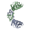
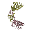
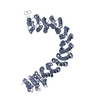
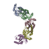
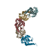
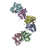
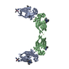
Structure of Cap5 from Lactococcus lactis
Components
SAVED domain-containing protein
Keywords
UNKNOWN FUNCTION / DNA nuclease SAVED Sensor Effector
Function / homology
HNH endonuclease / SMODS-associated and fused to various effectors / SMODS-associated and fused to various effectors sensor domain / HNH nuclease / metal ion binding / SMODS-associated and fused to various effectors domain-containing protein
A: SAVED domain-containing protein B: SAVED domain-containing protein C: SAVED domain-containing protein D: SAVED domain-containing protein hetero molecules
 Movie
Movie Controller


 Open data
 Basic information
 Components
 Keywords
 Function and homology information
 Lactococcus lactis subsp. cremoris IBB477 (lactic acid bacteria)
 X-RAY DIFFRACTION /
  Authors
 United States, 1items
  Citation
 Structure visualization
 Downloads & links
 Other downloads
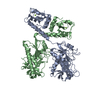

 PDBj
PDBj
 Assembly