+ Open data
- Basic information
| Entry | Database: PDB / ID: 7rw9 | ||||||
|---|---|---|---|---|---|---|---|
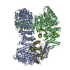
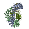
| Title | AP2 bound to heparin in the bowl conformation | ||||||
 Components Components |
| ||||||
 Keywords Keywords | ENDOCYTOSIS / AP2 / clathrin vesicle / lipid-binding / adaptor / membrane / transport | ||||||
| Function / homology |  Function and homology information Function and homology informationFormation of annular gap junctions / Gap junction degradation / LDL clearance / WNT5A-dependent internalization of FZD2, FZD5 and ROR2 / WNT5A-dependent internalization of FZD4 / Trafficking of GluR2-containing AMPA receptors / Retrograde neurotrophin signalling / VLDLR internalisation and degradation / clathrin adaptor complex / cardiac septum development ...Formation of annular gap junctions / Gap junction degradation / LDL clearance / WNT5A-dependent internalization of FZD2, FZD5 and ROR2 / WNT5A-dependent internalization of FZD4 / Trafficking of GluR2-containing AMPA receptors / Retrograde neurotrophin signalling / VLDLR internalisation and degradation / clathrin adaptor complex / cardiac septum development / Recycling pathway of L1 / extrinsic component of presynaptic endocytic zone membrane / regulation of vesicle size / postsynaptic endocytic zone / AP-2 adaptor complex / postsynaptic neurotransmitter receptor internalization / Cargo recognition for clathrin-mediated endocytosis / membrane coat / Clathrin-mediated endocytosis / clathrin coat assembly / positive regulation of synaptic vesicle endocytosis / clathrin adaptor activity / vesicle budding from membrane / clathrin-dependent endocytosis / MHC class II antigen presentation / signal sequence binding / coronary vasculature development / positive regulation of protein localization to membrane / neurotransmitter secretion / regulation of hematopoietic stem cell differentiation / aorta development / ventricular septum development / low-density lipoprotein particle receptor binding / clathrin binding / positive regulation of endocytosis / positive regulation of receptor internalization / synaptic vesicle endocytosis / negative regulation of protein localization to plasma membrane / vesicle-mediated transport / Neutrophil degranulation / secretory granule / intracellular protein transport / kidney development / kinase binding / cytoplasmic side of plasma membrane / disordered domain specific binding / synaptic vesicle / presynapse / heart development / protein-containing complex assembly / cytoplasmic vesicle / transmembrane transporter binding / postsynapse / protein domain specific binding / synapse / lipid binding / protein kinase binding / protein-containing complex binding / glutamatergic synapse / mitochondrion Similarity search - Function | ||||||
| Biological species |  | ||||||
| Method | ELECTRON MICROSCOPY / single particle reconstruction / cryo EM / Resolution: 3.9 Å | ||||||
 Authors Authors | Baker, R.W. / Hollopeter, G. / Partlow, E.A. | ||||||
| Funding support |  United States, 1items United States, 1items
| ||||||
 Citation Citation |  Journal: Nat Struct Mol Biol / Year: 2022 Journal: Nat Struct Mol Biol / Year: 2022Title: Structural basis of an endocytic checkpoint that primes the AP2 clathrin adaptor for cargo internalization. Authors: Edward A Partlow / Kevin S Cannon / Gunther Hollopeter / Richard W Baker /  Abstract: Clathrin-mediated endocytosis (CME) is the main route of internalization from the plasma membrane. It is known that the heterotetrameric AP2 clathrin adaptor must open to simultaneously engage ...Clathrin-mediated endocytosis (CME) is the main route of internalization from the plasma membrane. It is known that the heterotetrameric AP2 clathrin adaptor must open to simultaneously engage membrane and endocytic cargo, yet it is unclear how transmembrane cargos are captured to catalyze CME. Using cryogenic-electron microscopy, we discover a new way in which mouse AP2 can reorganize to expose membrane- and cargo-binding pockets, which is not observed in clathrin-coated structures. Instead, it is stimulated by endocytic pioneer proteins called muniscins, which do not enter vesicles. Muniscin-engaged AP2 is primed to rearrange into the vesicle-competent conformation on binding the tyrosine cargo internalization motif (YxxΦ). We propose adaptor priming as a checkpoint to ensure cargo internalization. | ||||||
| History |
|
- Structure visualization
| Structure viewer | Molecule:  Molmil Molmil Jmol/JSmol Jmol/JSmol |
|---|
- Downloads & links
- Download
| PDBx/mmCIF format |  7rw9.cif.gz 7rw9.cif.gz | 256 KB | Display |  PDBx/mmCIF format PDBx/mmCIF format |
|---|---|---|---|---|
| PDB format |  pdb7rw9.ent.gz pdb7rw9.ent.gz | 192.9 KB | Display |  PDB format PDB format |
| PDBx/mmJSON format |  7rw9.json.gz 7rw9.json.gz | Tree view |  PDBx/mmJSON format PDBx/mmJSON format | |
| Others |  Other downloads Other downloads |
-Validation report
| Summary document |  7rw9_validation.pdf.gz 7rw9_validation.pdf.gz | 1.3 MB | Display |  wwPDB validaton report wwPDB validaton report |
|---|---|---|---|---|
| Full document |  7rw9_full_validation.pdf.gz 7rw9_full_validation.pdf.gz | 1.3 MB | Display | |
| Data in XML |  7rw9_validation.xml.gz 7rw9_validation.xml.gz | 54.4 KB | Display | |
| Data in CIF |  7rw9_validation.cif.gz 7rw9_validation.cif.gz | 81.5 KB | Display | |
| Arichive directory |  https://data.pdbj.org/pub/pdb/validation_reports/rw/7rw9 https://data.pdbj.org/pub/pdb/validation_reports/rw/7rw9 ftp://data.pdbj.org/pub/pdb/validation_reports/rw/7rw9 ftp://data.pdbj.org/pub/pdb/validation_reports/rw/7rw9 | HTTPS FTP |
-Related structure data
| Related structure data |  24711MC  7rw8C  7rwaC  7rwbC  7rwcC C: citing same article ( M: map data used to model this data |
|---|---|
| Similar structure data | Similarity search - Function & homology  F&H Search F&H Search |
- Links
Links
- Assembly
| Deposited unit | 
|
|---|---|
| 1 |
|
- Components
| #1: Protein | Mass: 70573.320 Da / Num. of mol.: 1 Source method: isolated from a genetically manipulated source Details: Expressed as a C-terminal GST fusion and the tag was removed with Prescission protease Source: (gene. exp.)   |
|---|---|
| #2: Protein | Mass: 66953.195 Da / Num. of mol.: 1 Source method: isolated from a genetically manipulated source Source: (gene. exp.)   |
| #3: Protein | Mass: 49726.641 Da / Num. of mol.: 1 Source method: isolated from a genetically manipulated source Source: (gene. exp.)   |
| #4: Protein | Mass: 17038.688 Da / Num. of mol.: 1 Source method: isolated from a genetically manipulated source Source: (gene. exp.)   |
-Experimental details
-Experiment
| Experiment | Method: ELECTRON MICROSCOPY |
|---|---|
| EM experiment | Aggregation state: PARTICLE / 3D reconstruction method: single particle reconstruction |
- Sample preparation
| Component | Name: AP2 complex bound to heparin in the "bowl" conformation Type: COMPLEX / Entity ID: all / Source: RECOMBINANT | ||||||||||||||||||||
|---|---|---|---|---|---|---|---|---|---|---|---|---|---|---|---|---|---|---|---|---|---|
| Molecular weight | Experimental value: NO | ||||||||||||||||||||
| Source (natural) | Organism:  | ||||||||||||||||||||
| Source (recombinant) | Organism:  | ||||||||||||||||||||
| Buffer solution | pH: 7.4 | ||||||||||||||||||||
| Buffer component |
| ||||||||||||||||||||
| Specimen | Conc.: 2 mg/ml / Embedding applied: NO / Shadowing applied: NO / Staining applied: NO / Vitrification applied: YES Details: Heparin added at a 2-fold excess 20 mM HEPES pH 7.4, 150 mM NaCl, 1 mM DTT, 0.05% beta-OG | ||||||||||||||||||||
| Specimen support | Grid material: COPPER / Grid mesh size: 300 divisions/in. / Grid type: Quantifoil R1.2/1.3 | ||||||||||||||||||||
| Vitrification | Instrument: FEI VITROBOT MARK IV / Cryogen name: ETHANE-PROPANE / Humidity: 100 % / Chamber temperature: 277 K / Details: Blot force -10, 4 second blot, 4 uL sample |
- Electron microscopy imaging
| Experimental equipment |  Model: Talos Arctica / Image courtesy: FEI Company |
|---|---|
| Microscopy | Model: FEI TALOS ARCTICA |
| Electron gun | Electron source:  FIELD EMISSION GUN / Accelerating voltage: 200 kV / Illumination mode: FLOOD BEAM FIELD EMISSION GUN / Accelerating voltage: 200 kV / Illumination mode: FLOOD BEAM |
| Electron lens | Mode: BRIGHT FIELD / Nominal magnification: 45000 X / Nominal defocus max: 2500 nm / Nominal defocus min: 500 nm / Cs: 2.7 mm / C2 aperture diameter: 70 µm / Alignment procedure: COMA FREE |
| Specimen holder | Cryogen: NITROGEN |
| Image recording | Electron dose: 55 e/Å2 / Film or detector model: GATAN K3 BIOQUANTUM (6k x 4k) |
- Processing
| Software | Name: PHENIX / Version: 1.19.2_4158: / Classification: refinement | ||||||||||||||||||||||||||||||||
|---|---|---|---|---|---|---|---|---|---|---|---|---|---|---|---|---|---|---|---|---|---|---|---|---|---|---|---|---|---|---|---|---|---|
| EM software |
| ||||||||||||||||||||||||||||||||
| Image processing | Details: Counting Mode | ||||||||||||||||||||||||||||||||
| CTF correction | Type: PHASE FLIPPING AND AMPLITUDE CORRECTION | ||||||||||||||||||||||||||||||||
| Particle selection | Num. of particles selected: 805304 | ||||||||||||||||||||||||||||||||
| 3D reconstruction | Resolution: 3.9 Å / Resolution method: FSC 0.143 CUT-OFF / Num. of particles: 151164 / Symmetry type: POINT | ||||||||||||||||||||||||||||||||
| Atomic model building | Protocol: RIGID BODY FIT / Details: phenix.real_space_refine | ||||||||||||||||||||||||||||||||
| Atomic model building | PDB-ID: 2VGL Accession code: 2VGL / Source name: PDB / Type: experimental model | ||||||||||||||||||||||||||||||||
| Refine LS restraints |
|
 Movie
Movie Controller


 PDBj
PDBj


