 Movie
Movie Controller

[English] 日本語
 Yorodumi
- PDB-7rst: The Crystal Structure of Recombinant Chloroperoxidase Expressed i... -
+ Open data

- Basic information
| Entry | Database: PDB / ID: 7rst | ||||||
|---|---|---|---|---|---|---|---|
| Title | The Crystal Structure of Recombinant Chloroperoxidase Expressed in Aspergillus niger | ||||||
 Components Components | Chloroperoxidase | ||||||
 Keywords Keywords | OXIDOREDUCTASE / recombinant chloroperoxidase / Aspergillus niger | ||||||
| Function / homology |  Function and homology information Function and homology information | ||||||
| Biological species |  Leptoxyphium fumago (fungus) Leptoxyphium fumago (fungus) | ||||||
| Method |  X-RAY DIFFRACTION / SYNCHROTRON / MOLECULAR REPLACEMENT / Resolution: 1.69 Å X-RAY DIFFRACTION / SYNCHROTRON / MOLECULAR REPLACEMENT / Resolution: 1.69 Å | ||||||
 Authors Authors | Tang, X. / Venkadesh, S. / Zhou, J. / Rosen, B. / Wang, X. | ||||||
| Funding support |  United States, 1items United States, 1items
| ||||||
 Citation Citation | Journal: To Be Published Title: The Crystal Structure of Recombinant Chloroperoxidase Expressed in Aspergillus niger Authors: Tang, X. / Venkadesh, S. / Zhou, J. / Rosen, B. / Wang, X. | ||||||
| History |
|
- Structure visualization
| Structure viewer | Molecule: MolmilJmol/JSmol |
|---|
- Downloads & links
-Download
| PDBx/mmCIF format | 7rst.cif.gz | 95.7 KB | Display | PDBx/mmCIF format |
|---|---|---|---|---|
| PDB format | pdb7rst.ent.gz | 69.2 KB | Display | PDB format |
| PDBx/mmJSON format | 7rst.json.gz | Tree view | PDBx/mmJSON format | |
| Others |  Other downloads Other downloads |
-Validation report
| Arichive directory | https://data.pdbj.org/pub/pdb/validation_reports/rs/7rstftp://data.pdbj.org/pub/pdb/validation_reports/rs/7rst | HTTPS FTP |
|---|
-Related structure data
| Related structure data | 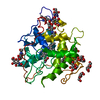 2cpoS S: Starting model for refinement |
|---|---|
| Similar structure data |
-Links
 PDBj
PDBj


- Assembly
| Deposited unit | 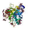
| ||||||||
|---|---|---|---|---|---|---|---|---|---|
| 1 |
| ||||||||
| Unit cell |
|
-Components
-Protein , 1 types, 1 molecules A
| #1: Protein | Mass: 32746.885 Da / Num. of mol.: 1 Source method: isolated from a genetically manipulated source Source: (gene. exp.) Leptoxyphium fumago (fungus) / Gene: CPO / Production host: |
|---|
-Sugars , 4 types, 16 molecules 

| #2: Polysaccharide | Source method: isolated from a genetically manipulated source #3: Polysaccharide | alpha-D-mannopyranose-(1-2)-alpha-D-mannopyranose | Source method: isolated from a genetically manipulated source #6: Sugar | ChemComp-NAG / |  Type: D-saccharide, beta linking / Mass: 221.208 Da / Num. of mol.: 1 Type: D-saccharide, beta linking / Mass: 221.208 Da / Num. of mol.: 1Source method: isolated from a genetically manipulated source Formula: C8H15NO6 #7: Sugar | ChemComp-MAN /  Type: D-saccharide, alpha linking / Mass: 180.156 Da / Num. of mol.: 12 Type: D-saccharide, alpha linking / Mass: 180.156 Da / Num. of mol.: 12Source method: isolated from a genetically manipulated source Formula: C6H12O6 |
|---|
-Non-polymers , 4 types, 480 molecules 


| #4: Chemical | ChemComp-MN /  Mass: 54.938 Da / Num. of mol.: 1 / Source method: obtained synthetically / Formula: Mn Mass: 54.938 Da / Num. of mol.: 1 / Source method: obtained synthetically / Formula: Mn | ||
|---|---|---|---|
| #5: Chemical | ChemComp-HEM /  Mass: 616.487 Da / Num. of mol.: 1 Mass: 616.487 Da / Num. of mol.: 1Source method: isolated from a genetically manipulated source Formula: C34H32FeN4O4 / Feature type: SUBJECT OF INVESTIGATION | ||
| #8: Chemical | ChemComp-IOD /  Mass: 126.904 Da / Num. of mol.: 10 / Source method: obtained synthetically / Formula: I Mass: 126.904 Da / Num. of mol.: 10 / Source method: obtained synthetically / Formula: I#9: Water | ChemComp-HOH / | Mass: 18.015 Da / Num. of mol.: 468 / Source method: isolated from a natural source / Formula: H2O |
-Details
| Has ligand of interest | Y |
|---|---|
| Has protein modification | Y |
-Experimental details
-Experiment
| Experiment | Method: X-RAY DIFFRACTION / Number of used crystals: 1 |
|---|
- Sample preparation
| Crystal | Density Matthews: 3.37 Å3/Da / Density % sol: 63.51 % |
|---|---|
| Crystal grow | Temperature: 293.15 K / Method: vapor diffusion, sitting drop / Details: 0.2M NaI, 20% polyethyleneglycol (PEG) 3350 |
-Data collection
| Diffraction | Mean temperature: 100 K / Serial crystal experiment: N |
|---|---|
| Diffraction source | Source: SYNCHROTRON / Site: ALS / Beamline: 8.2.2 / Wavelength: 0.9999 Å |
| Detector | Type: ADSC QUANTUM 315 / Detector: CCD / Date: Oct 19, 2016 |
| Radiation | Protocol: SINGLE WAVELENGTH / Monochromatic (M) / Laue (L): M / Scattering type: x-ray |
| Radiation wavelength | Wavelength: 0.9999 Å / Relative weight: 1 |
| Reflection | Resolution: 1.69→37.3 Å / Num. obs: 45316 / % possible obs: 90.5 % / Redundancy: 6.9 % / Rmerge(I) obs: 0.109 / Net I/σ(I): 9.4 |
| Reflection shell | Resolution: 1.69→1.78 Å / Rmerge(I) obs: 0.597 / Num. unique obs: 7220 |
- Processing
| Software |
| |||||||||||||||||||||||||||||||||||||||||||||||||||||||||||||||||||||||||||||||||||||||||||||||||||||||||||||||||||||||
|---|---|---|---|---|---|---|---|---|---|---|---|---|---|---|---|---|---|---|---|---|---|---|---|---|---|---|---|---|---|---|---|---|---|---|---|---|---|---|---|---|---|---|---|---|---|---|---|---|---|---|---|---|---|---|---|---|---|---|---|---|---|---|---|---|---|---|---|---|---|---|---|---|---|---|---|---|---|---|---|---|---|---|---|---|---|---|---|---|---|---|---|---|---|---|---|---|---|---|---|---|---|---|---|---|---|---|---|---|---|---|---|---|---|---|---|---|---|---|---|---|
| Refinement | Method to determine structure: MOLECULAR REPLACEMENT Starting model: 2CPO Resolution: 1.69→30.36 Å / SU ML: 0.21 / Cross valid method: THROUGHOUT / σ(F): 1.35 / Phase error: 18.62 / Stereochemistry target values: ML
| |||||||||||||||||||||||||||||||||||||||||||||||||||||||||||||||||||||||||||||||||||||||||||||||||||||||||||||||||||||||
| Solvent computation | Shrinkage radii: 0.9 Å / VDW probe radii: 1.11 Å / Solvent model: FLAT BULK SOLVENT MODEL | |||||||||||||||||||||||||||||||||||||||||||||||||||||||||||||||||||||||||||||||||||||||||||||||||||||||||||||||||||||||
| Displacement parameters | Biso max: 97.76 Å2 / Biso mean: 22.0758 Å2 / Biso min: 8.2 Å2 | |||||||||||||||||||||||||||||||||||||||||||||||||||||||||||||||||||||||||||||||||||||||||||||||||||||||||||||||||||||||
| Refinement step | Cycle: final / Resolution: 1.69→30.36 Å
| |||||||||||||||||||||||||||||||||||||||||||||||||||||||||||||||||||||||||||||||||||||||||||||||||||||||||||||||||||||||
| Refine LS restraints |
| |||||||||||||||||||||||||||||||||||||||||||||||||||||||||||||||||||||||||||||||||||||||||||||||||||||||||||||||||||||||
| LS refinement shell | Refine-ID: X-RAY DIFFRACTION / Rfactor Rfree error: 0 / Total num. of bins used: 16
|
