National Institutes of Health/National Institute of General Medical Sciences (NIH/NIGMS)
P41GM136508
United States
National Institutes of Health/National Institute of General Medical Sciences (NIH/NIGMS)
R35 GM127086
United States
National Institutes of Health/National Institute of General Medical Sciences (NIH/NIGMS)
R01GM124152
United States
Citation
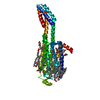
Journal: Proc Natl Acad Sci U S A / Year: 2021 Title: MicroED structure of the human adenosine receptor determined from a single nanocrystal in LCP. Authors: Michael W Martynowycz / Anna Shiriaeva / Xuanrui Ge / Johan Hattne / Brent L Nannenga / Vadim Cherezov / Tamir Gonen / Abstract: G protein-coupled receptors (GPCRs), or seven-transmembrane receptors, are a superfamily of membrane proteins that are critically important to physiological processes in the human body. Determining ...G protein-coupled receptors (GPCRs), or seven-transmembrane receptors, are a superfamily of membrane proteins that are critically important to physiological processes in the human body. Determining high-resolution structures of GPCRs without bound cognate signaling partners, such as a G protein, requires crystallization in lipidic cubic phase (LCP). GPCR crystals grown in LCP are often too small for traditional X-ray crystallography. These microcrystals are ideal for investigation by microcrystal electron diffraction (MicroED), but the gel-like nature of LCP makes traditional approaches to MicroED sample preparation insurmountable. Here, we show that the structure of a human A adenosine receptor can be determined by MicroED after converting the LCP into the sponge phase followed by focused ion-beam milling. We determined the structure of the A adenosine receptor to 2.8-Å resolution and resolved an antagonist in its orthosteric ligand-binding site, as well as four cholesterol molecules bound around the receptor. This study lays the groundwork for future structural studies of lipid-embedded membrane proteins by MicroED using single microcrystals that would be impossible with other crystallographic methods.
Cryogen: NITROGEN / Specimen holder model: FEI TITAN KRIOS AUTOGRID HOLDER / Temperature (max): 83 K / Temperature (min): 77 K
Image recording
Average exposure time: 3 sec. / Electron dose: 0.01 e/Å2 / Film or detector model: FEI CETA (4k x 4k) / Num. of diffraction images: 134 / Num. of grids imaged: 1
Image scans
Sampling size: 28 µm / Width: 2048 / Height: 2048
EM diffraction
Camera length: 1900 mm / Tilt angle list: -40, +40, 0.6
EM diffraction shell
Resolution: 37.91→2.79 Å / Fourier space coverage: 77 % / Multiplicity: 3.7 / Num. of structure factors: 10071 / Phase residual: 30 °
EM diffraction stats
Fourier space coverage: 77 % / High resolution: 2.79 Å / Num. of intensities measured: 37130 / Num. of structure factors: 10071 / Phase error: 30 ° / Phase error rejection criteria: none / Rmerge: 0.299
Diffraction
Serial crystal experiment: N
Detector
Detector: CMOS / Date: Sep 11, 2020
-
Processing
Software
Name
Classification
DIALS
datareduction
PHASER
phasing
PHENIX
refinement
EM software
ID
Name
Category
1
EPU
imageacquisition
6
Coot
modelfitting
8
PHENIX
modelrefinement
9
PHASER
molecularreplacement
11
DIALS
symmetrydetermination
13
PHENIX
3Dreconstruction
Image processing
Details: CetaD binned by 2
EM 3D crystal entity
∠α: 90 ° / ∠β: 90 ° / ∠γ: 90 ° / A: 40 Å / B: 180.5 Å / C: 139.7 Å / Space group name: C2221 / Space group num: 20
CTF correction
Type: NONE
3D reconstruction
Resolution: 2.79 Å / Resolution method: DIFFRACTION PATTERN/LAYERLINES / Algorithm: FOURIER SPACE / Symmetry type: 3D CRYSTAL
Atomic model building
B value: 43 / Protocol: RIGID BODY FIT / Space: RECIPROCAL / Target criteria: Maximum likelihood
In the structure databanks used in Yorodumi, some data are registered as the other names, "COVID-19 virus" and "2019-nCoV". Here are the details of the virus and the list of structure data.
Jan 31, 2019. EMDB accession codes are about to change! (news from PDBe EMDB page)
EMDB accession codes are about to change! (news from PDBe EMDB page)
The allocation of 4 digits for EMDB accession codes will soon come to an end. Whilst these codes will remain in use, new EMDB accession codes will include an additional digit and will expand incrementally as the available range of codes is exhausted. The current 4-digit format prefixed with “EMD-” (i.e. EMD-XXXX) will advance to a 5-digit format (i.e. EMD-XXXXX), and so on. It is currently estimated that the 4-digit codes will be depleted around Spring 2019, at which point the 5-digit format will come into force.
The EM Navigator/Yorodumi systems omit the EMD- prefix.
Related info.:Q: What is EMD? / ID/Accession-code notation in Yorodumi/EM Navigator
Yorodumi is a browser for structure data from EMDB, PDB, SASBDB, etc.
This page is also the successor to EM Navigator detail page, and also detail information page/front-end page for Omokage search.
The word "yorodu" (or yorozu) is an old Japanese word meaning "ten thousand". "mi" (miru) is to see.
Related info.:EMDB / PDB / SASBDB / Comparison of 3 databanks / Yorodumi Search / Aug 31, 2016. New EM Navigator & Yorodumi / Yorodumi Papers / Jmol/JSmol / Function and homology information / Changes in new EM Navigator and Yorodumi
 Movie
Movie Controller


 Open data
 Basic information
 Components
 Keywords
 Function and homology information
 Homo sapiens (human)
 MOLECULAR REPLACEMENT / cryo EM / Resolution: 2.79 Å
  Authors
 United States, 4items
  Citation
 Structure visualization
 Downloads & links
 Other downloads
 PDBj
PDBj


 Assembly


 Spodoptera frugiperda (fall armyworm) / References: UniProt: P29274, UniProt: P0ABE7
 Mass: 337.336 Da / Num. of mol.: 1 / Source method: obtained synthetically / Formula: C16H15N7O2 / Comment: antagonist*YM
 Mass: 386.654 Da / Num. of mol.: 4 / Source method: obtained synthetically / Formula: C27H46O
 Mass: 22.990 Da / Num. of mol.: 1 / Source method: obtained synthetically / Formula: Na
 Mass: 18.015 Da / Num. of mol.: 6 / Source method: isolated from a natural source / Formula: H2O
 Sample preparation
 Processing