Movie
Movie Controller


+ Open data

- Basic information
| Entry | Database: EMDB / ID: EMD-24551 | |||||||||||||||
|---|---|---|---|---|---|---|---|---|---|---|---|---|---|---|---|---|
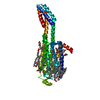
| Title | MicroED structure of the human adenosine receptor at 2.8A | |||||||||||||||
 Map data Map data | 2mFo-dFc map of A2A AR by MicroED | |||||||||||||||
 Sample Sample |
| |||||||||||||||
 Keywords Keywords | MEMBRANE PROTEIN | |||||||||||||||
| Function / homology |  Function and homology information Function and homology informationregulation of norepinephrine secretion / positive regulation of circadian sleep/wake cycle, sleep / Adenosine P1 receptors / positive regulation of acetylcholine secretion, neurotransmission / G protein-coupled adenosine receptor activity / response to purine-containing compound / G protein-coupled adenosine receptor signaling pathway / NGF-independant TRKA activation / Surfactant metabolism / type 5 metabotropic glutamate receptor binding ...regulation of norepinephrine secretion / positive regulation of circadian sleep/wake cycle, sleep / Adenosine P1 receptors / positive regulation of acetylcholine secretion, neurotransmission / G protein-coupled adenosine receptor activity / response to purine-containing compound / G protein-coupled adenosine receptor signaling pathway / NGF-independant TRKA activation / Surfactant metabolism / type 5 metabotropic glutamate receptor binding / negative regulation of vascular permeability / intermediate filament / presynaptic active zone / positive regulation of urine volume / response to caffeine / blood circulation / synaptic transmission, cholinergic / sensory perception / positive regulation of glutamate secretion / eating behavior / regulation of calcium ion transport / alpha-actinin binding / synaptic transmission, dopaminergic / membrane depolarization / asymmetric synapse / axolemma / cellular defense response / prepulse inhibition / phagocytosis / neuron projection morphogenesis / positive regulation of synaptic transmission, glutamatergic / astrocyte activation / presynaptic modulation of chemical synaptic transmission / excitatory postsynaptic potential / regulation of mitochondrial membrane potential / positive regulation of long-term synaptic potentiation / positive regulation of protein secretion / central nervous system development / synaptic transmission, glutamatergic / locomotory behavior / electron transport chain / vasodilation / blood coagulation / adenylate cyclase-modulating G protein-coupled receptor signaling pathway / cell-cell signaling / adenylate cyclase-activating G protein-coupled receptor signaling pathway / presynaptic membrane / negative regulation of neuron apoptotic process / G alpha (s) signalling events / phospholipase C-activating G protein-coupled receptor signaling pathway / positive regulation of ERK1 and ERK2 cascade / calmodulin binding / periplasmic space / electron transfer activity / postsynaptic membrane / response to xenobiotic stimulus / iron ion binding / inflammatory response / negative regulation of cell population proliferation / neuronal cell body / heme binding / apoptotic process / regulation of DNA-templated transcription / lipid binding / dendrite / protein-containing complex binding / glutamatergic synapse / enzyme binding / membrane / identical protein binding / plasma membrane Similarity search - Function | |||||||||||||||
| Biological species |  Homo sapiens (human) Homo sapiens (human) | |||||||||||||||
| Method | electron crystallography / cryo EM / Resolution: 2.79 Å | |||||||||||||||
 Authors Authors | Martynowycz MW / Shiriaeva A | |||||||||||||||
| Funding support |  United States, 4 items United States, 4 items
| |||||||||||||||
 Citation Citation | Journal: Proc Natl Acad Sci U S A / Year: 2021 Title: MicroED structure of the human adenosine receptor determined from a single nanocrystal in LCP. Authors: Michael W Martynowycz / Anna Shiriaeva / Xuanrui Ge / Johan Hattne / Brent L Nannenga / Vadim Cherezov / Tamir Gonen / Abstract: G protein-coupled receptors (GPCRs), or seven-transmembrane receptors, are a superfamily of membrane proteins that are critically important to physiological processes in the human body. Determining ...G protein-coupled receptors (GPCRs), or seven-transmembrane receptors, are a superfamily of membrane proteins that are critically important to physiological processes in the human body. Determining high-resolution structures of GPCRs without bound cognate signaling partners, such as a G protein, requires crystallization in lipidic cubic phase (LCP). GPCR crystals grown in LCP are often too small for traditional X-ray crystallography. These microcrystals are ideal for investigation by microcrystal electron diffraction (MicroED), but the gel-like nature of LCP makes traditional approaches to MicroED sample preparation insurmountable. Here, we show that the structure of a human A adenosine receptor can be determined by MicroED after converting the LCP into the sponge phase followed by focused ion-beam milling. We determined the structure of the A adenosine receptor to 2.8-Å resolution and resolved an antagonist in its orthosteric ligand-binding site, as well as four cholesterol molecules bound around the receptor. This study lays the groundwork for future structural studies of lipid-embedded membrane proteins by MicroED using single microcrystals that would be impossible with other crystallographic methods. | |||||||||||||||
| History |
|
- Structure visualization
| Movie |
Movie viewer |
|---|---|
| Structure viewer | EM map: SurfViewMolmilJmol/JSmol |
| Supplemental images |

- Downloads & links
-EMDB archive
| Map data | emd_24551.map.gz | 4.3 MB | EMDB map data format | |
|---|---|---|---|---|
| Header (meta data) | emd-24551-v30.xmlemd-24551.xml | 16.9 KB 16.9 KB | Display Display | EMDB header |
| Images |  emd_24551.png emd_24551.png | 35.2 KB | ||
| Filedesc metadata | emd-24551.cif.gz | 6.4 KB | ||
| Filedesc structureFactors | emd_24551_sf.cif.gz | 294.8 KB | ||
| Archive directory |  http://ftp.pdbj.org/pub/emdb/structures/EMD-24551ftp://ftp.pdbj.org/pub/emdb/structures/EMD-24551 http://ftp.pdbj.org/pub/emdb/structures/EMD-24551ftp://ftp.pdbj.org/pub/emdb/structures/EMD-24551 | HTTPS FTP |
-Related structure data
| Related structure data |  7rm5MC M: atomic model generated by this map C: citing same article ( |
|---|---|
| Similar structure data |
-Links
| EMDB pages | EMDB (EBI/PDBe) / EMDataResource |
|---|---|
| Related items in Molecule of the Month |


-Map
| File | Download / File: emd_24551.map.gz / Format: CCP4 / Size: 4.8 MB / Type: IMAGE STORED AS FLOATING POINT NUMBER (4 BYTES) | ||||||||||||||||||||||||||||||||||||||||||||||||||||||||||||||||||||
|---|---|---|---|---|---|---|---|---|---|---|---|---|---|---|---|---|---|---|---|---|---|---|---|---|---|---|---|---|---|---|---|---|---|---|---|---|---|---|---|---|---|---|---|---|---|---|---|---|---|---|---|---|---|---|---|---|---|---|---|---|---|---|---|---|---|---|---|---|---|
| Annotation | 2mFo-dFc map of A2A AR by MicroED | ||||||||||||||||||||||||||||||||||||||||||||||||||||||||||||||||||||
| Projections & slices | Image control
Images are generated by Spider. generated in cubic-lattice coordinate | ||||||||||||||||||||||||||||||||||||||||||||||||||||||||||||||||||||
| Voxel size | X: 0.667 Å / Y: 0.669 Å / Z: 0.698 Å | ||||||||||||||||||||||||||||||||||||||||||||||||||||||||||||||||||||
| Density |
| ||||||||||||||||||||||||||||||||||||||||||||||||||||||||||||||||||||
| Symmetry | Space group: 20 | ||||||||||||||||||||||||||||||||||||||||||||||||||||||||||||||||||||
| Details | EMDB XML:
CCP4 map header:
| ||||||||||||||||||||||||||||||||||||||||||||||||||||||||||||||||||||
 X (Sec.)
 Y (Row.)
 Z (Col.)


-Supplemental data
- Sample components
-Entire : Adenosine receptor A2a/Soluble cytochrome b562 chimera
| Entire | Name: Adenosine receptor A2a/Soluble cytochrome b562 chimera |
|---|---|
| Components |
|
-Supramolecule #1: Adenosine receptor A2a/Soluble cytochrome b562 chimera
| Supramolecule | Name: Adenosine receptor A2a/Soluble cytochrome b562 chimera type: complex / ID: 1 / Parent: 0 / Macromolecule list: #1 |
|---|---|
| Source (natural) | Organism: Homo sapiens (human) |
| Molecular weight | Theoretical: 58.72 KDa |
-Macromolecule #1: Adenosine receptor A2a/Soluble cytochrome b562 chimera
| Macromolecule | Name: Adenosine receptor A2a/Soluble cytochrome b562 chimera type: protein_or_peptide / ID: 1 / Number of copies: 1 / Enantiomer: LEVO |
|---|---|
| Source (natural) | Organism: Homo sapiens (human) |
| Molecular weight | Theoretical: 49.974281 KDa |
| Recombinant expression | Organism:   Spodoptera frugiperda (fall armyworm) Spodoptera frugiperda (fall armyworm) |
| Sequence | String: MKTIIALSYI FCLVFADYKD DDDGAPPIMG SSVYITVELA IAVLAILGNV LVCWAVWLNS NLQNVTNYFV VSLAAADIAV GVLAIPFAI TISTGFCAAC HGCLFIACFV LVLTQSSIFS LLAIAIDRYI AIRIPLRYNG LVTGTRAKGI IAICWVLSFA I GLTPMLGW ...String: MKTIIALSYI FCLVFADYKD DDDGAPPIMG SSVYITVELA IAVLAILGNV LVCWAVWLNS NLQNVTNYFV VSLAAADIAV GVLAIPFAI TISTGFCAAC HGCLFIACFV LVLTQSSIFS LLAIAIDRYI AIRIPLRYNG LVTGTRAKGI IAICWVLSFA I GLTPMLGW NNCGQPKEGK NHSQGCGEGQ VACLFEDVVP MNYMVYFNFF ACVLVPLLLM LGVYLRIFLA ARRQLADLED NW ETLNDNL KVIEKADNAA QVKDALTKMR AAALDAQKAT PPKLEDKSPD SPEMKDFRHG FDILVGQIDD ALKLANEGKV KEA QAAAEQ LKTTRNAYIQ KYLERARSTL QKEVHAAKSL AIIVGLFALC WLPLHIINCF TFFCPDCSHA PLWLMYLAIV LSHT NSVVN PFIYAYRIRE FRQTFRKIIR SHVLRQQEPF KAHHHHHHHH HH UniProtKB: Adenosine receptor A2a, Soluble cytochrome b562, Adenosine receptor A2a |
-Macromolecule #2: 4-{2-[(7-amino-2-furan-2-yl[1,2,4]triazolo[1,5-a][1,3,5]triazin-5...
| Macromolecule | Name: 4-{2-[(7-amino-2-furan-2-yl[1,2,4]triazolo[1,5-a][1,3,5]triazin-5-yl)amino]ethyl}phenol type: ligand / ID: 2 / Number of copies: 1 / Formula: ZMA |
|---|---|
| Molecular weight | Theoretical: 337.336 Da |
| Chemical component information |  ChemComp-ZMA: |
-Macromolecule #3: CHOLESTEROL
| Macromolecule | Name: CHOLESTEROL / type: ligand / ID: 3 / Number of copies: 4 / Formula: CLR |
|---|---|
| Molecular weight | Theoretical: 386.654 Da |
| Chemical component information |  ChemComp-CLR: |
-Macromolecule #4: SODIUM ION
| Macromolecule | Name: SODIUM ION / type: ligand / ID: 4 / Number of copies: 1 |
|---|---|
| Molecular weight | Theoretical: 22.99 Da |
-Macromolecule #5: water
| Macromolecule | Name: water / type: ligand / ID: 5 / Number of copies: 6 / Formula: HOH |
|---|---|
| Molecular weight | Theoretical: 18.015 Da |
| Chemical component information |  ChemComp-HOH: |
-Experimental details
-Structure determination
| Method | cryo EM |
|---|---|
 Processing Processing | electron crystallography |
| Aggregation state | 3D array |
-Sample preparation
| Concentration | 10 mg/mL |
|---|---|
| Buffer | pH: 5 Details: 25-28% (v/v) PEG 400, 0.04-0.06M sodium thiocyanate, 2% (v/v) 2,5-hexanediol, 100mM sodium citrate, pH 5.0, Lipid Cubic Phase (LCP), temperature 293K |
| Grid | Model: Quantifoil R2/2 / Material: COPPER / Mesh: 200 / Support film - Material: CARBON / Support film - topology: HOLEY ARRAY / Pretreatment - Type: GLOW DISCHARGE / Pretreatment - Time: 30 sec. |
| Vitrification | Cryogen name: ETHANE / Chamber humidity: 95 % / Chamber temperature: 298 K / Instrument: LEICA EM GP |
- Electron microscopy
| Microscope | FEI TITAN KRIOS |
|---|---|
| Temperature | Min: 77.0 K / Max: 83.0 K |
| Image recording | Film or detector model: FEI CETA (4k x 4k) / Digitization - Dimensions - Width: 2048 pixel / Digitization - Dimensions - Height: 2048 pixel / Number grids imaged: 1 / Number diffraction images: 134 / Average exposure time: 3.0 sec. / Average electron dose: 0.01 e/Å2 |
| Electron beam | Acceleration voltage: 300 kV / Electron source:  FIELD EMISSION GUN FIELD EMISSION GUN |
| Electron optics | C2 aperture diameter: 50.0 µm / Illumination mode: FLOOD BEAM / Imaging mode: DIFFRACTION / Cs: 2.7 mm / Nominal defocus max: 0.0 µm / Nominal defocus min: 0.0 µm / Camera length: 1900 mm |
| Sample stage | Specimen holder model: FEI TITAN KRIOS AUTOGRID HOLDER / Cooling holder cryogen: NITROGEN / Tilt angle: -40.0, 40.0, 0.6 |
| Experimental equipment |  Model: Titan Krios / Image courtesy: FEI Company |
+Image processing
-Atomic model buiding 1
| Initial model | PDB ID: Chain - Chain ID: A / Chain - Source name: PDB / Chain - Initial model type: experimental model |
|---|---|
| Refinement | Space: RECIPROCAL / Protocol: RIGID BODY FIT / Overall B value: 43 / Target criteria: Maximum likelihood |
| Output model | PDB-7rm5: |