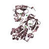
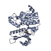
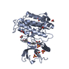
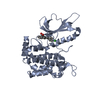
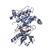
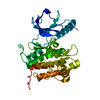
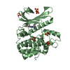
regulation of clathrin-dependent endocytosis / clathrin complex / AP-2 adaptor complex binding / Cargo recognition for clathrin-mediated endocytosis / Clathrin-mediated endocytosis / Notch binding / clathrin-coated vesicle / positive regulation of Notch signaling pathway / presynaptic endocytosis / cell leading edge ...regulation of clathrin-dependent endocytosis / clathrin complex / AP-2 adaptor complex binding / Cargo recognition for clathrin-mediated endocytosis / Clathrin-mediated endocytosis / Notch binding / clathrin-coated vesicle / positive regulation of Notch signaling pathway / presynaptic endocytosis / cell leading edge / clathrin-coated pit / calyx of Held / terminal bouton / regulation of protein localization / presynapse / protein phosphorylation / non-specific serine/threonine protein kinase / protein stabilization / protein serine kinase activity / protein serine/threonine kinase activity / ATP binding / plasma membrane / cytosol Similarity search - Function
: / Serine/threonine-protein kinase, active site / Serine/Threonine protein kinases active-site signature. / Protein kinase domain / Serine/Threonine protein kinases, catalytic domain / Protein kinase domain profile. / Protein kinase domain / Protein kinase-like domain superfamily Similarity search - Domain/homology
Mass: 18.015 Da / Num. of mol.: 217 / Source method: isolated from a natural source / Formula: H2O
Has ligand of interest
Y
-
Experimental details
-
Experiment
Experiment
Method: X-RAY DIFFRACTION / Number of used crystals: 1
-
Sample preparation
Crystal
Density Matthews: 2.92 Å3/Da / Density % sol: 57.94 %
Crystal grow
Temperature: 295 K / Method: vapor diffusion, hanging drop / pH: 5.5 Details: 0.9 M ammonium sulfate, 0.14 M sodium chloride, 0.1 M Bis-Tris pH 5.5, and 1% PEG 3350
-
Data collection
Diffraction
Mean temperature: 100 K / Serial crystal experiment: N
Protocol: SINGLE WAVELENGTH / Monochromatic (M) / Laue (L): M / Scattering type: x-ray
Radiation wavelength
Wavelength: 1 Å / Relative weight: 1
Reflection
Resolution: 2.12→58.44 Å / Num. obs: 43896 / % possible obs: 92.7 % / Redundancy: 9.6 % / Biso Wilson estimate: 58.46 Å2 / Rmerge(I) obs: 0.058 / Net I/σ(I): 18.4
Reflection shell
Resolution: 2.12→2.21 Å / Redundancy: 10.5 % / Rmerge(I) obs: 1.387 / Mean I/σ(I) obs: 1.5 / Num. unique obs: 2196 / % possible all: 48.7
-
Processing
Software
Name
Version
Classification
BUSTER
refinement
SCALA
3.3.16
datascaling
PDB_EXTRACT
3.27
dataextraction
XDS
datareduction
PHASER
phasing
Refinement
Method to determine structure: MOLECULAR REPLACEMENT Starting model: In house model Resolution: 2.124→21.24 Å / Cor.coef. Fo:Fc: 0.948 / Cor.coef. Fo:Fc free: 0.949 / SU R Cruickshank DPI: 0.459 / Cross valid method: THROUGHOUT / σ(F): 0 / SU R Blow DPI: 0.232 / SU Rfree Blow DPI: 0.178 / SU Rfree Cruickshank DPI: 0.177 Details: HYDROGENS WERE FULLY REFINED WITH ZERO OCCUPANCY AT NUCLEAR POSITION. REFINEMENT NOTES. NUMBER OF REFINEMENT NOTES : 1 NOTE 1 : IDEAL-DIST CONTACT TERM CONTACT SETUP. ALL ATOMS HAVE CCP4 ATOM TYPE FROM LIBRARY
In the structure databanks used in Yorodumi, some data are registered as the other names, "COVID-19 virus" and "2019-nCoV". Here are the details of the virus and the list of structure data.
Jan 31, 2019. EMDB accession codes are about to change! (news from PDBe EMDB page)
EMDB accession codes are about to change! (news from PDBe EMDB page)
The allocation of 4 digits for EMDB accession codes will soon come to an end. Whilst these codes will remain in use, new EMDB accession codes will include an additional digit and will expand incrementally as the available range of codes is exhausted. The current 4-digit format prefixed with “EMD-” (i.e. EMD-XXXX) will advance to a 5-digit format (i.e. EMD-XXXXX), and so on. It is currently estimated that the 4-digit codes will be depleted around Spring 2019, at which point the 5-digit format will come into force.
The EM Navigator/Yorodumi systems omit the EMD- prefix.
Related info.:Q: What is EMD? / ID/Accession-code notation in Yorodumi/EM Navigator
Yorodumi is a browser for structure data from EMDB, PDB, SASBDB, etc.
This page is also the successor to EM Navigator detail page, and also detail information page/front-end page for Omokage search.
The word "yorodu" (or yorozu) is an old Japanese word meaning "ten thousand". "mi" (miru) is to see.
Related info.:EMDB / PDB / SASBDB / Comparison of 3 databanks / Yorodumi Search / Aug 31, 2016. New EM Navigator & Yorodumi / Yorodumi Papers / Jmol/JSmol / Function and homology information / Changes in new EM Navigator and Yorodumi
 Movie
Movie Controller

 Yorodumi
 Open data
 Basic information
 Components
 Keywords
 Function and homology information
 X-RAY DIFFRACTION /
  Authors
 Citation
 Structure visualization
 Downloads & links
 Other downloads
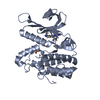


 PDBj
PDBj


 Assembly

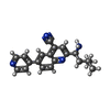
 Mass: 316.400 Da / Num. of mol.: 2 / Source method: obtained synthetically / Formula: C20H20N4 / Feature type: SUBJECT OF INVESTIGATION
 Mass: 96.063 Da / Num. of mol.: 6 / Source method: obtained synthetically / Formula: SO4
 Mass: 18.015 Da / Num. of mol.: 217 / Source method: isolated from a natural source / Formula: H2O
 Sample preparation
 / Beamline: 17-ID / Wavelength: 1 Å
 Processing