 Movie
Movie Controller

[English] 日本語
 Yorodumi
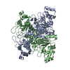
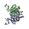
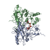
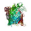
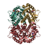
- PDB-7rh8: Crystal structure of Fur1p from Candida albicans, in complex with UTP -
+ Open data

- Basic information
| Entry | Database: PDB / ID: 7rh8 | ||||||
|---|---|---|---|---|---|---|---|
| Title | Crystal structure of Fur1p from Candida albicans, in complex with UTP | ||||||
 Components Components | Uracil phosphoribosyltransferase | ||||||
 Keywords Keywords | TRANSFERASE / fur1 / drug target / structural genomics / CSGID / Center for Structural Genomics of Infectious Diseases / NIAID / national institute of allergy and infectious diseases | ||||||
| Function / homology | pyrimidine-containing compound salvage / Uracil phosphoribosyltransferase / uracil phosphoribosyltransferase activity / fungal biofilm matrix / Phosphoribosyltransferase-like / Phosphoribosyltransferase domain / URIDINE 5'-TRIPHOSPHATE / Uracil phosphoribosyltransferase Function and homology information Function and homology information | ||||||
| Biological species |  Candida albicans (yeast) Candida albicans (yeast) | ||||||
| Method |  X-RAY DIFFRACTION / SYNCHROTRON / MOLECULAR REPLACEMENT / Resolution: 1.69 Å X-RAY DIFFRACTION / SYNCHROTRON / MOLECULAR REPLACEMENT / Resolution: 1.69 Å | ||||||
 Authors Authors | Stogios, P.J. / Skarina, T. / Kim, Y. / Di Leo, R. / Savchenko, A. / Joachimiak, A. / Satchell, K.J.F. / Center for Structural Genomics of Infectious Diseases (CSGID) | ||||||
| Funding support |  United States, 1items United States, 1items
| ||||||
 Citation Citation | Journal: To Be Published Title: Crystal structure of Fur1p from Candida albicans, in complex with UTP Authors: Stogios, P.J. | ||||||
| History |
|
- Structure visualization
| Structure viewer | Molecule: MolmilJmol/JSmol |
|---|
- Downloads & links
-Download
| PDBx/mmCIF format | 7rh8.cif.gz | 121.6 KB | Display | PDBx/mmCIF format |
|---|---|---|---|---|
| PDB format | pdb7rh8.ent.gz | 82 KB | Display | PDB format |
| PDBx/mmJSON format | 7rh8.json.gz | Tree view | PDBx/mmJSON format | |
| Others |  Other downloads Other downloads |
-Validation report
| Arichive directory | https://data.pdbj.org/pub/pdb/validation_reports/rh/7rh8ftp://data.pdbj.org/pub/pdb/validation_reports/rh/7rh8 | HTTPS FTP |
|---|
-Related structure data
| Related structure data |  1jlsS S: Starting model for refinement |
|---|---|
| Similar structure data | |
| Other databases |


-Links
 PDBj
PDBj
- Assembly
| Deposited unit | 
| |||||||||||||||||||||||||||
|---|---|---|---|---|---|---|---|---|---|---|---|---|---|---|---|---|---|---|---|---|---|---|---|---|---|---|---|---|
| 1 | 
| |||||||||||||||||||||||||||
| Unit cell |
| |||||||||||||||||||||||||||
| Components on special symmetry positions |
|
-Components
| #1: Protein | Mass: 24506.744 Da / Num. of mol.: 1 Source method: isolated from a genetically manipulated source Source: (gene. exp.) Candida albicans (strain SC5314 / ATCC MYA-2876) (yeast)Strain: SC5314 / ATCC MYA-2876 / Gene: FUR1, orf19.2640, CAALFM_C503390CA / Plasmid: pMCSG53 / Production host:  |
|---|---|
| #2: Chemical | ChemComp-UTP /   Mass: 484.141 Da / Num. of mol.: 1 / Source method: obtained synthetically / Formula: C9H15N2O15P3 / Feature type: SUBJECT OF INVESTIGATION / Comment: UTP*YM Mass: 484.141 Da / Num. of mol.: 1 / Source method: obtained synthetically / Formula: C9H15N2O15P3 / Feature type: SUBJECT OF INVESTIGATION / Comment: UTP*YM |
| #3: Water | ChemComp-HOH /  Mass: 18.015 Da / Num. of mol.: 323 / Source method: isolated from a natural source / Formula: H2O Mass: 18.015 Da / Num. of mol.: 323 / Source method: isolated from a natural source / Formula: H2O |
| Has ligand of interest | Y |
-Experimental details
-Experiment
| Experiment | Method: X-RAY DIFFRACTION / Number of used crystals: 1 |
|---|
- Sample preparation
| Crystal | Density Matthews: 3.2 Å3/Da / Density % sol: 61.57 % |
|---|---|
| Crystal grow | Temperature: 298 K / Method: vapor diffusion, sitting drop / pH: 5.6 Details: 25% (w/v) PEG3350, 0.2 M NaCl, 0.1 M sodium citrate pH 5.6 |
-Data collection
| Diffraction | Mean temperature: 100 K / Serial crystal experiment: N |
|---|---|
| Diffraction source | Source: SYNCHROTRON / Site: APS / Beamline: 19-ID / Wavelength: 0.97869 Å |
| Detector | Type: DECTRIS PILATUS3 6M / Detector: PIXEL / Date: Apr 3, 2021 |
| Radiation | Protocol: SINGLE WAVELENGTH / Monochromatic (M) / Laue (L): M / Scattering type: x-ray |
| Radiation wavelength | Wavelength: 0.97869 Å / Relative weight: 1 |
| Reflection | Resolution: 1.69→50 Å / Num. obs: 35544 / % possible obs: 98 % / Redundancy: 13.4 % / Biso Wilson estimate: 22 Å2 / CC1/2: 1 / Rmerge(I) obs: 0.08 / Rpim(I) all: 0.022 / Net I/σ(I): 29.73 |
| Reflection shell | Resolution: 1.7→1.73 Å / Rmerge(I) obs: 0.844 / Mean I/σ(I) obs: 1.6 / Num. unique obs: 1496 / CC1/2: 0.949 / Rpim(I) all: 0.262 / % possible all: 84.3 |
- Processing
| Software |
| ||||||||||||||||||||||||||||||||||||||||||||||||||||||||||||||||||||||||||||||||||||||||||||||||||||
|---|---|---|---|---|---|---|---|---|---|---|---|---|---|---|---|---|---|---|---|---|---|---|---|---|---|---|---|---|---|---|---|---|---|---|---|---|---|---|---|---|---|---|---|---|---|---|---|---|---|---|---|---|---|---|---|---|---|---|---|---|---|---|---|---|---|---|---|---|---|---|---|---|---|---|---|---|---|---|---|---|---|---|---|---|---|---|---|---|---|---|---|---|---|---|---|---|---|---|---|---|---|
| Refinement | Method to determine structure: MOLECULAR REPLACEMENT Starting model: 1JLS Resolution: 1.69→47.78 Å / SU ML: 0.1571 / Cross valid method: FREE R-VALUE / σ(F): 1.35 / Phase error: 18.381 Stereochemistry target values: GeoStd + Monomer Library + CDL v1.2
| ||||||||||||||||||||||||||||||||||||||||||||||||||||||||||||||||||||||||||||||||||||||||||||||||||||
| Solvent computation | Shrinkage radii: 0.9 Å / VDW probe radii: 1.11 Å / Solvent model: FLAT BULK SOLVENT MODEL | ||||||||||||||||||||||||||||||||||||||||||||||||||||||||||||||||||||||||||||||||||||||||||||||||||||
| Displacement parameters | Biso mean: 32.1 Å2 | ||||||||||||||||||||||||||||||||||||||||||||||||||||||||||||||||||||||||||||||||||||||||||||||||||||
| Refinement step | Cycle: LAST / Resolution: 1.69→47.78 Å
| ||||||||||||||||||||||||||||||||||||||||||||||||||||||||||||||||||||||||||||||||||||||||||||||||||||
| Refine LS restraints |
| ||||||||||||||||||||||||||||||||||||||||||||||||||||||||||||||||||||||||||||||||||||||||||||||||||||
| LS refinement shell |
| ||||||||||||||||||||||||||||||||||||||||||||||||||||||||||||||||||||||||||||||||||||||||||||||||||||
| Refinement TLS params. | Method: refined / Refine-ID: X-RAY DIFFRACTION
| ||||||||||||||||||||||||||||||||||||||||||||||||||||||||||||||||||||||||||||||||||||||||||||||||||||
| Refinement TLS group | Refine-ID: X-RAY DIFFRACTION / Auth asym-ID: A / Label asym-ID: A
|
