 Movie
Movie Controller

[English] 日本語
 Yorodumi
- PDB-7rgv: Structure of Caulobacter crescentus Suppressor of copper sensitiv... -
+ Open data

- Basic information
| Entry | Database: PDB / ID: 7rgv | ||||||
|---|---|---|---|---|---|---|---|
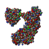
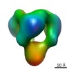
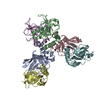
| Title | Structure of Caulobacter crescentus Suppressor of copper sensitivity protein C | ||||||
 Components Components | Thioredoxin domain-containing protein | ||||||
 Keywords Keywords | OXIDOREDUCTASE / thioredoxin-like / ScsC / isomerase / periplasmic | ||||||
| Function / homology |  Function and homology information Function and homology information | ||||||
| Biological species |  Caulobacter vibrioides (bacteria) Caulobacter vibrioides (bacteria) | ||||||
| Method |  X-RAY DIFFRACTION / SYNCHROTRON / MOLECULAR REPLACEMENT / Resolution: 2.63 Å X-RAY DIFFRACTION / SYNCHROTRON / MOLECULAR REPLACEMENT / Resolution: 2.63 Å | ||||||
 Authors Authors | Petit, G.A. / Martin, J.L. / Gulbis, J.M. | ||||||
| Funding support |  Australia, 1items Australia, 1items
| ||||||
 Citation Citation | Journal: Acta Crystallogr D Struct Biol / Year: 2022 Title: The suppressor of copper sensitivity protein C from Caulobacter crescentus is a trimeric disulfide isomerase that binds copper(I) with subpicomolar affinity. Authors: Petit, G.A. / Hong, Y. / Djoko, K.Y. / Whitten, A.E. / Furlong, E.J. / McCoy, A.J. / Gulbis, J.M. / Totsika, M. / Martin, J.L. / Halili, M.A. | ||||||
| History |
|
- Structure visualization
| Structure viewer | Molecule: MolmilJmol/JSmol |
|---|
- Downloads & links
-Download
| PDBx/mmCIF format | 7rgv.cif.gz | 161.4 KB | Display | PDBx/mmCIF format |
|---|---|---|---|---|
| PDB format | pdb7rgv.ent.gz | 109.3 KB | Display | PDB format |
| PDBx/mmJSON format | 7rgv.json.gz | Tree view | PDBx/mmJSON format | |
| Others |  Other downloads Other downloads |
-Validation report
| Arichive directory | https://data.pdbj.org/pub/pdb/validation_reports/rg/7rgvftp://data.pdbj.org/pub/pdb/validation_reports/rg/7rgv | HTTPS FTP |
|---|
-Related structure data
| Related structure data |  4vxwS S: Starting model for refinement |
|---|---|
| Similar structure data | |
| Other databases |

-Links
 PDBj
PDBj


- Assembly
| Deposited unit | 
| ||||||||||||
|---|---|---|---|---|---|---|---|---|---|---|---|---|---|
| 1 | 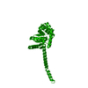
| ||||||||||||
| Unit cell |
|
-Components
| #1: Protein | Mass: 25166.631 Da / Num. of mol.: 1 / Mutation: C2S Source method: isolated from a genetically manipulated source Details: Linker and TEV scar not visible in the electron densities hence not modelled Source: (gene. exp.) Caulobacter vibrioides (strain ATCC 19089 / CB15) (bacteria)Strain: ATCC 19089 / CB15 / Gene: CC_1879 / Production host: |
|---|---|
| #2: Water | ChemComp-HOH /  Mass: 18.015 Da / Num. of mol.: 3 / Source method: isolated from a natural source / Formula: H2O Mass: 18.015 Da / Num. of mol.: 3 / Source method: isolated from a natural source / Formula: H2O |
| Has protein modification | Y |
-Experimental details
-Experiment
| Experiment | Method: X-RAY DIFFRACTION / Number of used crystals: 1 |
|---|
- Sample preparation
| Crystal | Density Matthews: 3.51 Å3/Da / Density % sol: 65 % / Description: thick hexagonal crystalline rod |
|---|---|
| Crystal grow | Temperature: 277.15 K / Method: vapor diffusion, hanging drop / pH: 7.25 Details: Protein reduced with DTT and purified in 25 mM HEPES pH 7.5, 150 mM NaCl. Protein concentrated to 130 mg/ml. 1ul of protein solution mixed with 1ul of crystallant solution: 39% MPD, 200 mM ...Details: Protein reduced with DTT and purified in 25 mM HEPES pH 7.5, 150 mM NaCl. Protein concentrated to 130 mg/ml. 1ul of protein solution mixed with 1ul of crystallant solution: 39% MPD, 200 mM NaAcetate, 20 mM CaCl2 pH 7.25 |
-Data collection
| Diffraction | Mean temperature: 100 K / Serial crystal experiment: N |
|---|---|
| Diffraction source | Source: SYNCHROTRON / Site: Australian Synchrotron / Beamline: MX2 / Wavelength: 0.9536 Å |
| Detector | Type: DECTRIS EIGER X 16M / Detector: PIXEL / Date: Feb 21, 2021 |
| Radiation | Protocol: SINGLE WAVELENGTH / Monochromatic (M) / Laue (L): M / Scattering type: x-ray |
| Radiation wavelength | Wavelength: 0.9536 Å / Relative weight: 1 |
| Reflection | Resolution: 2.63→49.34 Å / Num. obs: 10854 / % possible obs: 98.75 % / Redundancy: 12 % / Biso Wilson estimate: 90.52 Å2 / CC1/2: 1 / CC star: 1 / Rmerge(I) obs: 0.052 / Rpim(I) all: 0.016 / Rrim(I) all: 0.054 / Net I/σ(I): 22.51 |
| Reflection shell | Resolution: 2.63→2.74 Å / Redundancy: 12.3 % / Rmerge(I) obs: 1.46 / Mean I/σ(I) obs: 1.85 / Num. unique obs: 1045 / CC1/2: 0.961 / CC star: 0.99 / Rpim(I) all: 0.431 / Rrim(I) all: 1.52 / % possible all: 94.76 |
- Processing
| Software |
| |||||||||||||||||||||||||||||||||||||||||||||||||||||||||||||||||||||||||||
|---|---|---|---|---|---|---|---|---|---|---|---|---|---|---|---|---|---|---|---|---|---|---|---|---|---|---|---|---|---|---|---|---|---|---|---|---|---|---|---|---|---|---|---|---|---|---|---|---|---|---|---|---|---|---|---|---|---|---|---|---|---|---|---|---|---|---|---|---|---|---|---|---|---|---|---|---|
| Refinement | Method to determine structure: MOLECULAR REPLACEMENT Starting model: 4VXW res 50-224 chain A Resolution: 2.63→49.34 Å / SU ML: 0.3672 / Cross valid method: FREE R-VALUE / σ(F): 1.36 / Phase error: 40.1233 Stereochemistry target values: GeoStd + Monomer Library + CDL v1.2 Details: Cycle of automated refinement with Phenix + manual refinemetn with Coot. No real space refinement with Phenix, 2 TLS groups 2-63, 64-223. Secondary structure restraint to keep helices in ...Details: Cycle of automated refinement with Phenix + manual refinemetn with Coot. No real space refinement with Phenix, 2 TLS groups 2-63, 64-223. Secondary structure restraint to keep helices in place. Structure has very few crystal contact+ large solvent channel and is expected to be dynamic hence the very high B-factors
| |||||||||||||||||||||||||||||||||||||||||||||||||||||||||||||||||||||||||||
| Solvent computation | Shrinkage radii: 0.9 Å / VDW probe radii: 1.11 Å / Solvent model: FLAT BULK SOLVENT MODEL | |||||||||||||||||||||||||||||||||||||||||||||||||||||||||||||||||||||||||||
| Displacement parameters | Biso mean: 122.3 Å2 | |||||||||||||||||||||||||||||||||||||||||||||||||||||||||||||||||||||||||||
| Refinement step | Cycle: LAST / Resolution: 2.63→49.34 Å
| |||||||||||||||||||||||||||||||||||||||||||||||||||||||||||||||||||||||||||
| Refine LS restraints |
| |||||||||||||||||||||||||||||||||||||||||||||||||||||||||||||||||||||||||||
| LS refinement shell |
| |||||||||||||||||||||||||||||||||||||||||||||||||||||||||||||||||||||||||||
| Refinement TLS params. | Method: refined / Refine-ID: X-RAY DIFFRACTION
| |||||||||||||||||||||||||||||||||||||||||||||||||||||||||||||||||||||||||||
| Refinement TLS group | Refine-ID: X-RAY DIFFRACTION / Auth asym-ID: A / Label asym-ID: A
|
