 Movie
Movie Controller

[English] 日本語
 Yorodumi
- PDB-7rgc: Crystal structure of triosephosphate isomerase from Candidatus Ab... -
+ Open data

- Basic information
| Entry | Database: PDB / ID: 7rgc | ||||||
|---|---|---|---|---|---|---|---|
| Title | Crystal structure of triosephosphate isomerase from Candidatus Absconditabacteria (Sr1) bacterium | ||||||
 Components Components | Triosephosphate isomerase | ||||||
 Keywords Keywords | ISOMERASE / TIM / triosephosphate isomerase / glycolysis / gluconeogenesis | ||||||
| Function / homology |  Function and homology information Function and homology informationtriose-phosphate isomerase / triose-phosphate isomerase activity / glycerol catabolic process / glyceraldehyde-3-phosphate biosynthetic process / gluconeogenesis / glycolytic process / cytosol Similarity search - Function | ||||||
| Biological species |  candidate division SR1 bacterium RAAC1_SR1_1 (bacteria) candidate division SR1 bacterium RAAC1_SR1_1 (bacteria) | ||||||
| Method |  X-RAY DIFFRACTION / SYNCHROTRON / MOLECULAR REPLACEMENT / Resolution: 2.09 Å X-RAY DIFFRACTION / SYNCHROTRON / MOLECULAR REPLACEMENT / Resolution: 2.09 Å | ||||||
 Authors Authors | Vickers, C.J. / Compton, J. / Fraga, D. / Patrick, W.M. | ||||||
| Funding support |  New Zealand, 1items New Zealand, 1items
| ||||||
 Citation Citation | Journal: To Be Published Title: Structure of Sr1TPI - Candidatus Absconditabacteria bacterium triosephoshate isomerase Authors: Vickers, C.J. / Fraga, D. / Compton, J. / Patrick, W.M. | ||||||
| History |
|
- Structure visualization
| Structure viewer | Molecule: MolmilJmol/JSmol |
|---|
- Downloads & links
-Download
| PDBx/mmCIF format | 7rgc.cif.gz | 125.4 KB | Display | PDBx/mmCIF format |
|---|---|---|---|---|
| PDB format | pdb7rgc.ent.gz | 78.2 KB | Display | PDB format |
| PDBx/mmJSON format | 7rgc.json.gz | Tree view | PDBx/mmJSON format | |
| Others |  Other downloads Other downloads |
-Validation report
| Summary document | 7rgc_validation.pdf.gz | 440.1 KB | Display | wwPDB validaton report |
|---|---|---|---|---|
| Full document | 7rgc_full_validation.pdf.gz | 444.2 KB | Display | |
| Data in XML | 7rgc_validation.xml.gz | 19.1 KB | Display | |
| Data in CIF | 7rgc_validation.cif.gz | 26.5 KB | Display | |
| Arichive directory | https://data.pdbj.org/pub/pdb/validation_reports/rg/7rgcftp://data.pdbj.org/pub/pdb/validation_reports/rg/7rgc | HTTPS FTP |
-Related structure data
| Related structure data | 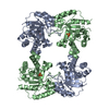 1b9bS S: Starting model for refinement |
|---|---|
| Similar structure data |
-Links
 PDBj
PDBj
- Assembly
| Deposited unit | 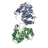
| ||||||||||||
|---|---|---|---|---|---|---|---|---|---|---|---|---|---|
| 1 | 
| ||||||||||||
| Unit cell |
|
-Components
| #1: Protein | Mass: 27606.562 Da / Num. of mol.: 2 Source method: isolated from a genetically manipulated source Source: (gene. exp.) candidate division SR1 bacterium RAAC1_SR1_1 (bacteria)Gene: tpiA, P148_SR1C00001G0257 / Production host: #2: Water | ChemComp-HOH / |  Mass: 18.015 Da / Num. of mol.: 112 / Source method: isolated from a natural source / Formula: H2O Mass: 18.015 Da / Num. of mol.: 112 / Source method: isolated from a natural source / Formula: H2OHas protein modification | Y | |
|---|
-Experimental details
-Experiment
| Experiment | Method: X-RAY DIFFRACTION / Number of used crystals: 1 |
|---|
- Sample preparation
| Crystal | Density Matthews: 2.39 Å3/Da / Density % sol: 48.49 % |
|---|---|
| Crystal grow | Temperature: 291.15 K / Method: vapor diffusion, hanging drop / Details: 2.3 M DL-Malic acid pH 7.0 |
-Data collection
| Diffraction | Mean temperature: 100 K / Serial crystal experiment: N |
|---|---|
| Diffraction source | Source: SYNCHROTRON / Site: Australian Synchrotron  / Beamline: MX2 / Wavelength: 0.95372 Å / Beamline: MX2 / Wavelength: 0.95372 Å |
| Detector | Type: ADSC QUANTUM 315r / Detector: CCD / Date: Feb 12, 2021 |
| Radiation | Protocol: SINGLE WAVELENGTH / Monochromatic (M) / Laue (L): M / Scattering type: x-ray |
| Radiation wavelength | Wavelength: 0.95372 Å / Relative weight: 1 |
| Reflection | Resolution: 2.09→45.82 Å / Num. obs: 30881 / % possible obs: 99.2 % / Redundancy: 20.3 % / Biso Wilson estimate: 31.73 Å2 / CC1/2: 0.999 / Net I/σ(I): 12.74 |
| Reflection shell | Resolution: 2.09→2.22 Å / Num. unique obs: 4732 / CC1/2: 0.83 |
- Processing
| Software |
| |||||||||||||||||||||||||||||||||||||||||||||||||||||||||||||||||||||||||||||||||||||||||||||||||||||||||
|---|---|---|---|---|---|---|---|---|---|---|---|---|---|---|---|---|---|---|---|---|---|---|---|---|---|---|---|---|---|---|---|---|---|---|---|---|---|---|---|---|---|---|---|---|---|---|---|---|---|---|---|---|---|---|---|---|---|---|---|---|---|---|---|---|---|---|---|---|---|---|---|---|---|---|---|---|---|---|---|---|---|---|---|---|---|---|---|---|---|---|---|---|---|---|---|---|---|---|---|---|---|---|---|---|---|---|
| Refinement | Method to determine structure: MOLECULAR REPLACEMENT Starting model: 1b9b Resolution: 2.09→44 Å / SU ML: 0.2863 / Cross valid method: FREE R-VALUE / σ(F): 1.33 / Phase error: 39.405 Stereochemistry target values: GeoStd + Monomer Library + CDL v1.2
| |||||||||||||||||||||||||||||||||||||||||||||||||||||||||||||||||||||||||||||||||||||||||||||||||||||||||
| Solvent computation | Shrinkage radii: 0.9 Å / VDW probe radii: 1.11 Å / Solvent model: FLAT BULK SOLVENT MODEL | |||||||||||||||||||||||||||||||||||||||||||||||||||||||||||||||||||||||||||||||||||||||||||||||||||||||||
| Displacement parameters | Biso mean: 47.73 Å2 | |||||||||||||||||||||||||||||||||||||||||||||||||||||||||||||||||||||||||||||||||||||||||||||||||||||||||
| Refinement step | Cycle: LAST / Resolution: 2.09→44 Å
| |||||||||||||||||||||||||||||||||||||||||||||||||||||||||||||||||||||||||||||||||||||||||||||||||||||||||
| Refine LS restraints |
| |||||||||||||||||||||||||||||||||||||||||||||||||||||||||||||||||||||||||||||||||||||||||||||||||||||||||
| LS refinement shell |
|
