 Movie
Movie Controller

[English] 日本語
 Yorodumi
- PDB-7rfq: STRUCTURE OF BACTERIAL SYLF DOMAIN CONTAINING PROTEIN, BETA CELL ... -
+ Open data

- Basic information
| Entry | Database: PDB / ID: 7rfq | ||||||
|---|---|---|---|---|---|---|---|
| Title | STRUCTURE OF BACTERIAL SYLF DOMAIN CONTAINING PROTEIN, BETA CELL EXPANSION FACTOR A (BEFA) | ||||||
 Components Components | BETA CELL EXPANSION FACTOR A (BEFA) | ||||||
 Keywords Keywords | SIGNALING PROTEIN / SYLF DOMAIN / BACTERIAL PROTEIN / DEVELOPMENTAL REGULATOR / BETA CELL EXPANSION FACTOR | ||||||
| Function / homology | host cellular component / extracellular region / CITRIC ACID / Beta cell expansion factor A Function and homology information Function and homology information | ||||||
| Biological species |  Aeromonas veronii (bacteria) Aeromonas veronii (bacteria) | ||||||
| Method |  X-RAY DIFFRACTION / SYNCHROTRON / SIRAS / Resolution: 1.27 Å X-RAY DIFFRACTION / SYNCHROTRON / SIRAS / Resolution: 1.27 Å | ||||||
 Authors Authors | Sweeney, E.G. | ||||||
| Funding support | 1items
| ||||||
 Citation Citation | Journal: Cell Metab. / Year: 2022 Title: BefA, a microbiota-secreted membrane disrupter, disseminates to the pancreas and increases beta cell mass. Authors: Hill, J.H. / Massaquoi, M.S. / Sweeney, E.G. / Wall, E.S. / Jahl, P. / Bell, R. / Kallio, K. / Derrick, D. / Murtaugh, L.C. / Parthasarathy, R. / Remington, S.J. / Round, J.L. / Guillemin, K. #1: Journal: Elife / Year: 2016 Title: A conserved bacterial protein induces pancreatic beta cell expansion during zebrafish development. Authors: Hill, J.H. / Franzosa, E.A. / Huttenhower, C. / Guillemin, K. | ||||||
| History |
|
- Structure visualization
| Structure viewer | Molecule: MolmilJmol/JSmol |
|---|
- Downloads & links
-Download
| PDBx/mmCIF format | 7rfq.cif.gz | 152.4 KB | Display | PDBx/mmCIF format |
|---|---|---|---|---|
| PDB format | pdb7rfq.ent.gz | 121.4 KB | Display | PDB format |
| PDBx/mmJSON format | 7rfq.json.gz | Tree view | PDBx/mmJSON format | |
| Others |  Other downloads Other downloads |
-Validation report
| Arichive directory | https://data.pdbj.org/pub/pdb/validation_reports/rf/7rfqftp://data.pdbj.org/pub/pdb/validation_reports/rf/7rfq | HTTPS FTP |
|---|
-Related structure data
| Similar structure data |
|---|
-Links
 PDBj
PDBj- Assembly
| Deposited unit | 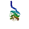
| ||||||||||||
|---|---|---|---|---|---|---|---|---|---|---|---|---|---|
| 1 |
| ||||||||||||
| Unit cell |
| ||||||||||||
| Components on special symmetry positions |
|
-Components
| #1: Protein | Mass: 29838.846 Da / Num. of mol.: 1 Source method: isolated from a genetically manipulated source Source: (gene. exp.) Aeromonas veronii (bacteria) / Gene: AMS64_07270 / Production host: | ||||
|---|---|---|---|---|---|
| #2: Chemical |   Mass: 192.124 Da / Num. of mol.: 2 / Source method: obtained synthetically / Formula: C6H8O7 Mass: 192.124 Da / Num. of mol.: 2 / Source method: obtained synthetically / Formula: C6H8O7#3: Water | ChemComp-HOH / |  Mass: 18.015 Da / Num. of mol.: 213 / Source method: isolated from a natural source / Formula: H2O Mass: 18.015 Da / Num. of mol.: 213 / Source method: isolated from a natural source / Formula: H2OHas ligand of interest | N | |
-Experimental details
-Experiment
| Experiment | Method: X-RAY DIFFRACTION / Number of used crystals: 1 |
|---|
- Sample preparation
| Crystal | Density Matthews: 2.01 Å3/Da / Density % sol: 38.66 % / Description: diamond-like, embedded in sticky precipitant |
|---|---|
| Crystal grow | Temperature: 295 K / Method: vapor diffusion, hanging drop / pH: 3.5 Details: 25 % PEG 3350, 100 mM CITRIC ACID PH 3.5, 1 mM TCEP, 39 mM O-PHOSPHO-L-SERINE OR 39 mM SERINE, VAPOR |
-Data collection
| Diffraction | Mean temperature: 80 K / Serial crystal experiment: N | |||||||||
|---|---|---|---|---|---|---|---|---|---|---|
| Diffraction source | Source: SYNCHROTRON / Site: ALS  / Beamline: 5.0.2 / Wavelength: 1.0, 0.9795 / Beamline: 5.0.2 / Wavelength: 1.0, 0.9795 | |||||||||
| Detector | Type: DECTRIS PILATUS3 S 6M / Detector: PIXEL / Date: Jul 16, 2016 | |||||||||
| Radiation | Protocol: SINGLE WAVELENGTH / Monochromatic (M) / Laue (L): M / Scattering type: x-ray | |||||||||
| Radiation wavelength |
| |||||||||
| Reflection | Resolution: 1.27→37.35 Å / Num. all: 62064 / Num. obs: 62064 / % possible obs: 100 % / Redundancy: 10 % / Rpim(I) all: 0.054 / Rrim(I) all: 0.177 / Rsym value: 0.168 / Net I/av σ(I): 2.4 / Net I/σ(I): 8.7 / Num. measured all: 621902 | |||||||||
| Reflection shell | Resolution: 1.27→1.32 Å / Redundancy: 11.5 % / Rmerge(I) obs: 0.114 / Mean I/σ(I) obs: 5 / Num. measured all: 23389 / Num. unique obs: 2032 / Rpim(I) all: 0.035 / Rrim(I) all: 0.119 / Rsym value: 0.114 / Net I/σ(I) obs: 17.2 / % possible all: 99.9 |
- Processing
| Software |
| ||||||||||||||||||||||||||||||||||||||||||||||||||||||||||||||||||||||||||||||||||||||||||||||||||||||
|---|---|---|---|---|---|---|---|---|---|---|---|---|---|---|---|---|---|---|---|---|---|---|---|---|---|---|---|---|---|---|---|---|---|---|---|---|---|---|---|---|---|---|---|---|---|---|---|---|---|---|---|---|---|---|---|---|---|---|---|---|---|---|---|---|---|---|---|---|---|---|---|---|---|---|---|---|---|---|---|---|---|---|---|---|---|---|---|---|---|---|---|---|---|---|---|---|---|---|---|---|---|---|---|
| Refinement | Method to determine structure: SIRAS / Resolution: 1.27→37.35 Å / SU ML: 0.1 / Cross valid method: THROUGHOUT / σ(F): 1.34 / Phase error: 13.54
| ||||||||||||||||||||||||||||||||||||||||||||||||||||||||||||||||||||||||||||||||||||||||||||||||||||||
| Solvent computation | Shrinkage radii: 0.9 Å / VDW probe radii: 1.11 Å | ||||||||||||||||||||||||||||||||||||||||||||||||||||||||||||||||||||||||||||||||||||||||||||||||||||||
| Displacement parameters | Biso max: 66.51 Å2 / Biso mean: 18.8979 Å2 / Biso min: 5.01 Å2 | ||||||||||||||||||||||||||||||||||||||||||||||||||||||||||||||||||||||||||||||||||||||||||||||||||||||
| Refinement step | Cycle: final / Resolution: 1.27→37.35 Å
| ||||||||||||||||||||||||||||||||||||||||||||||||||||||||||||||||||||||||||||||||||||||||||||||||||||||
| LS refinement shell | Refine-ID: X-RAY DIFFRACTION / Rfactor Rfree error: 0
|
