 Movie
Movie Controller


+ Open data

- Basic information
| Entry | Database: PDB / ID: 7rdw | ||||||
|---|---|---|---|---|---|---|---|
| Title | Crystal Structure of FH1 Fab bound to HXb2 HIV-1 gp120 core | ||||||
 Components Components |
| ||||||
 Keywords Keywords | IMMUNE SYSTEM / FH1 / VH1-2*02 / VRC01 / Antibody / HIV-1 | ||||||
| Function / homology | HIV Envelope Protein Gp120; Chain G / Human immunodeficiency virus 1, Gp160, envelope glycoprotein / Beta Complex / Immunoglobulins / Immunoglobulin-like / Sandwich / Mainly Beta / AMMONIUM ION Function and homology information Function and homology information | ||||||
| Biological species |   Human immunodeficiency virus 1 Human immunodeficiency virus 1 Homo sapiens (human) Homo sapiens (human) | ||||||
| Method |  X-RAY DIFFRACTION / SYNCHROTRON / MOLECULAR REPLACEMENT / Resolution: 3.55 Å X-RAY DIFFRACTION / SYNCHROTRON / MOLECULAR REPLACEMENT / Resolution: 3.55 Å | ||||||
 Authors Authors | Weidle, C. / Pancera, M. | ||||||
| Funding support |  United States, 1items United States, 1items
| ||||||
 Citation Citation | Journal: Sci Adv / Year: 2022 Title: Characterization of a vaccine-elicited human antibody with sequence homology to VRC01-class antibodies that binds the C1C2 gp120 domain. Authors: Gray, M.D. / Feng, J. / Weidle, C.E. / Cohen, K.W. / Ballweber-Fleming, L. / MacCamy, A.J. / Huynh, C.N. / Trichka, J.J. / Montefiori, D. / Ferrari, G. / Pancera, M. / McElrath, M.J. / Stamatatos, L. | ||||||
| History |
|
- Structure visualization
| Structure viewer | Molecule: MolmilJmol/JSmol |
|---|
- Downloads & links
-Download
| PDBx/mmCIF format | 7rdw.cif.gz | 626.8 KB | Display | PDBx/mmCIF format |
|---|---|---|---|---|
| PDB format | pdb7rdw.ent.gz | 458.1 KB | Display | PDB format |
| PDBx/mmJSON format | 7rdw.json.gz | Tree view | PDBx/mmJSON format | |
| Others |  Other downloads Other downloads |
-Validation report
| Arichive directory | https://data.pdbj.org/pub/pdb/validation_reports/rd/7rdwftp://data.pdbj.org/pub/pdb/validation_reports/rd/7rdw | HTTPS FTP |
|---|
-Related structure data
| Related structure data | 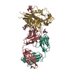 6mftS S: Starting model for refinement |
|---|---|
| Similar structure data |
-Links
 PDBj
PDBj


- Assembly
| Deposited unit | 
| ||||||||||||
|---|---|---|---|---|---|---|---|---|---|---|---|---|---|
| 1 | 
| ||||||||||||
| 2 | 
| ||||||||||||
| 3 | 
| ||||||||||||
| 4 | 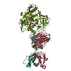
| ||||||||||||
| Unit cell |
|
-Components
-Protein , 1 types, 4 molecules CDMN
| #1: Protein | Mass: 41336.422 Da / Num. of mol.: 4 / Fragment: HXB2 gp120 core Source method: isolated from a genetically manipulated source Source: (gene. exp.) Human immunodeficiency virus 1 / Cell line (production host): 293 / Production host: Homo sapiens (human) |
|---|
-Antibody , 3 types, 8 molecules GUHQILRV
| #2: Antibody | Mass: 24472.371 Da / Num. of mol.: 2 Source method: isolated from a genetically manipulated source Source: (gene. exp.) Homo sapiens (human) / Production host: Homo sapiens (human)#3: Antibody | Mass: 24489.400 Da / Num. of mol.: 2 Source method: isolated from a genetically manipulated source Source: (gene. exp.) Homo sapiens (human) / Cell line (production host): 293 / Production host: Homo sapiens (human)#4: Antibody | Mass: 23185.830 Da / Num. of mol.: 4 Source method: isolated from a genetically manipulated source Source: (gene. exp.) Homo sapiens (human) / Production host: Homo sapiens (human) |
|---|
-Sugars , 2 types, 32 molecules 
| #5: Polysaccharide | beta-D-mannopyranose-(1-4)-2-acetamido-2-deoxy-beta-D-glucopyranose-(1-4)-2-acetamido-2-deoxy-beta- ...beta-D-mannopyranose-(1-4)-2-acetamido-2-deoxy-beta-D-glucopyranose-(1-4)-2-acetamido-2-deoxy-beta-D-glucopyranose Source method: isolated from a genetically manipulated source #6: Sugar | ChemComp-NAG /  Type: D-saccharide, beta linking / Mass: 221.208 Da / Num. of mol.: 28 Type: D-saccharide, beta linking / Mass: 221.208 Da / Num. of mol.: 28Source method: isolated from a genetically manipulated source Formula: C8H15NO6 |
|---|
-Non-polymers , 4 types, 102 molecules 


| #7: Chemical | ChemComp-NH4 /  Mass: 18.038 Da / Num. of mol.: 4 / Source method: obtained synthetically / Formula: H4N Mass: 18.038 Da / Num. of mol.: 4 / Source method: obtained synthetically / Formula: H4N#8: Chemical | ChemComp-EDO /  Mass: 62.068 Da / Num. of mol.: 6 / Source method: obtained synthetically / Formula: C2H6O2 Mass: 62.068 Da / Num. of mol.: 6 / Source method: obtained synthetically / Formula: C2H6O2#9: Chemical |  Mass: 96.063 Da / Num. of mol.: 3 / Source method: obtained synthetically / Formula: SO4 Mass: 96.063 Da / Num. of mol.: 3 / Source method: obtained synthetically / Formula: SO4#10: Water | ChemComp-HOH / | Mass: 18.015 Da / Num. of mol.: 89 / Source method: isolated from a natural source / Formula: H2O |
|---|
-Details
| Has ligand of interest | N |
|---|---|
| Has protein modification | Y |
-Experimental details
-Experiment
| Experiment | Method: X-RAY DIFFRACTION / Number of used crystals: 1 |
|---|
- Sample preparation
| Crystal | Density Matthews: 2.92 Å3/Da / Density % sol: 57.91 % |
|---|---|
| Crystal grow | Temperature: 293 K / Method: vapor diffusion, hanging drop / Details: 0.1 M NH4SO4, 20% PEG 1500, 0.1M Tris pH 7.5 |
-Data collection
| Diffraction | Mean temperature: 100 K / Serial crystal experiment: N |
|---|---|
| Diffraction source | Source: SYNCHROTRON / Site: ALS / Beamline: 5.0.2 / Wavelength: 1 Å |
| Detector | Type: DECTRIS PILATUS3 6M / Detector: PIXEL / Date: Nov 1, 2018 |
| Radiation | Protocol: SINGLE WAVELENGTH / Monochromatic (M) / Laue (L): M / Scattering type: x-ray |
| Radiation wavelength | Wavelength: 1 Å / Relative weight: 1 |
| Reflection | Resolution: 3.5→50.18 Å / Num. obs: 36171 / % possible obs: 92.5 % / Redundancy: 1.6 % / Biso Wilson estimate: 62.36 Å2 / CC1/2: 0.95 / Net I/σ(I): 5.29 |
| Reflection shell | Resolution: 3.5→3.56 Å / Num. unique obs: 1577 / CC1/2: 0.712 / % possible all: 69.3 |
- Processing
| Software |
| ||||||||||||||||||||||||||||||||||||||||||||||||||||||||||||||||||||||||||||||||||||||||||||||||||
|---|---|---|---|---|---|---|---|---|---|---|---|---|---|---|---|---|---|---|---|---|---|---|---|---|---|---|---|---|---|---|---|---|---|---|---|---|---|---|---|---|---|---|---|---|---|---|---|---|---|---|---|---|---|---|---|---|---|---|---|---|---|---|---|---|---|---|---|---|---|---|---|---|---|---|---|---|---|---|---|---|---|---|---|---|---|---|---|---|---|---|---|---|---|---|---|---|---|---|---|
| Refinement | Method to determine structure: MOLECULAR REPLACEMENT Starting model: 6MFT Resolution: 3.55→50.18 Å / SU ML: 0.4539 / Cross valid method: FREE R-VALUE / σ(F): 1.97 / Phase error: 31.1724 Stereochemistry target values: GeoStd + Monomer Library + CDL v1.2
| ||||||||||||||||||||||||||||||||||||||||||||||||||||||||||||||||||||||||||||||||||||||||||||||||||
| Solvent computation | Shrinkage radii: 0.9 Å / VDW probe radii: 1.11 Å / Solvent model: FLAT BULK SOLVENT MODEL | ||||||||||||||||||||||||||||||||||||||||||||||||||||||||||||||||||||||||||||||||||||||||||||||||||
| Displacement parameters | Biso mean: 70.99 Å2 | ||||||||||||||||||||||||||||||||||||||||||||||||||||||||||||||||||||||||||||||||||||||||||||||||||
| Refinement step | Cycle: LAST / Resolution: 3.55→50.18 Å
| ||||||||||||||||||||||||||||||||||||||||||||||||||||||||||||||||||||||||||||||||||||||||||||||||||
| Refine LS restraints |
| ||||||||||||||||||||||||||||||||||||||||||||||||||||||||||||||||||||||||||||||||||||||||||||||||||
| LS refinement shell |
|
