+ Open data
- Basic information
| Entry | Database: PDB / ID: 7ra7 | |||||||||
|---|---|---|---|---|---|---|---|---|---|---|
| Title | Crystal structure of rabbit anti-HIV Fab 11A | |||||||||
 Components Components |
| |||||||||
 Keywords Keywords | IMMUNE SYSTEM / antibody antigen neutralizing antibody HIV | |||||||||
| Function / homology | Immunoglobulins / Immunoglobulin-like / Sandwich / Mainly Beta Function and homology information Function and homology information | |||||||||
| Biological species |  | |||||||||
| Method |  X-RAY DIFFRACTION / X-RAY DIFFRACTION /  SYNCHROTRON / SYNCHROTRON /  MOLECULAR REPLACEMENT / Resolution: 2.2 Å MOLECULAR REPLACEMENT / Resolution: 2.2 Å | |||||||||
 Authors Authors | Oyen, D. / Wilson, I.A. | |||||||||
| Funding support |  United States, 2items United States, 2items
| |||||||||
 Citation Citation |  Journal: To Be Published Journal: To Be PublishedTitle: Limited breadth of anti-HIV Env glycan hole antibodies is further hindered by strain-specific peptide interactions Authors: Ozorowski, G. / Cottrell, C.A. / Oyen, D. / de Val, N. / Copps, J. / Scaring, N. / Polveroni, T.M. / Wilson, I.A. / Ward, A.B. #1:  Journal: Immunity / Year: 2018 Journal: Immunity / Year: 2018Title: Electron-Microscopy-Based Epitope Mapping Defines Specificities of Polyclonal Antibodies Elicited during HIV-1 BG505 Envelope Trimer Immunization. Authors: Matteo Bianchi / Hannah L Turner / Bartek Nogal / Christopher A Cottrell / David Oyen / Matthias Pauthner / Raiza Bastidas / Rebecca Nedellec / Laura E McCoy / Ian A Wilson / Dennis R Burton ...Authors: Matteo Bianchi / Hannah L Turner / Bartek Nogal / Christopher A Cottrell / David Oyen / Matthias Pauthner / Raiza Bastidas / Rebecca Nedellec / Laura E McCoy / Ian A Wilson / Dennis R Burton / Andrew B Ward / Lars Hangartner /   Abstract: Characterizing polyclonal antibody responses via currently available methods is inherently complex and difficult. Mapping epitopes in an immune response is typically incomplete, which creates a ...Characterizing polyclonal antibody responses via currently available methods is inherently complex and difficult. Mapping epitopes in an immune response is typically incomplete, which creates a barrier to fully understanding the humoral response to antigens and hinders rational vaccine design efforts. Here, we describe a method of characterizing polyclonal responses by using electron microscopy, and we applied this method to the immunization of rabbits with an HIV-1 envelope glycoprotein vaccine candidate, BG505 SOSIP.664. We detected known epitopes within the polyclonal sera and revealed how antibody responses evolved during the prime-boosting strategy to ultimately result in a neutralizing antibody response. We uncovered previously unidentified epitopes, including an epitope proximal to one recognized by human broadly neutralizing antibodies as well as potentially distracting non-neutralizing epitopes. Our method provides an efficient and semiquantitative map of epitopes that are targeted in a polyclonal antibody response and should be of widespread utility in vaccine and infection studies. #2:  Journal: Cell Rep / Year: 2016 Journal: Cell Rep / Year: 2016Title: Holes in the Glycan Shield of the Native HIV Envelope Are a Target of Trimer-Elicited Neutralizing Antibodies. Authors: Laura E McCoy / Marit J van Gils / Gabriel Ozorowski / Terrence Messmer / Bryan Briney / James E Voss / Daniel W Kulp / Matthew S Macauley / Devin Sok / Matthias Pauthner / Sergey Menis / ...Authors: Laura E McCoy / Marit J van Gils / Gabriel Ozorowski / Terrence Messmer / Bryan Briney / James E Voss / Daniel W Kulp / Matthew S Macauley / Devin Sok / Matthias Pauthner / Sergey Menis / Christopher A Cottrell / Jonathan L Torres / Jessica Hsueh / William R Schief / Ian A Wilson / Andrew B Ward / Rogier W Sanders / Dennis R Burton /    Abstract: A major advance in the search for an HIV vaccine has been the development of a near-native Envelope trimer (BG505 SOSIP.664) that can induce robust autologous Tier 2 neutralization. Here, potently ...A major advance in the search for an HIV vaccine has been the development of a near-native Envelope trimer (BG505 SOSIP.664) that can induce robust autologous Tier 2 neutralization. Here, potently neutralizing monoclonal antibodies (nAbs) from rabbits immunized with BG505 SOSIP.664 are shown to recognize an immunodominant region of gp120 centered on residue 241. Residue 241 occupies a hole in the glycan defenses of the BG505 isolate, with fewer than 3% of global isolates lacking a glycan site at this position. However, at least one conserved glycan site is missing in 89% of viruses, suggesting the presence of glycan holes in most HIV isolates. Serum evidence is consistent with targeting of holes in natural infection. The immunogenic nature of breaches in the glycan shield has been under-appreciated in previous attempts to understand autologous neutralizing antibody responses and has important potential consequences for HIV vaccine design. | |||||||||
| History |
|
- Structure visualization
| Structure viewer | Molecule:  Molmil Molmil Jmol/JSmol Jmol/JSmol |
|---|
- Downloads & links
- Download
| PDBx/mmCIF format |  7ra7.cif.gz 7ra7.cif.gz | 382.6 KB | Display |  PDBx/mmCIF format PDBx/mmCIF format |
|---|---|---|---|---|
| PDB format |  pdb7ra7.ent.gz pdb7ra7.ent.gz | 251.8 KB | Display |  PDB format PDB format |
| PDBx/mmJSON format |  7ra7.json.gz 7ra7.json.gz | Tree view |  PDBx/mmJSON format PDBx/mmJSON format | |
| Others |  Other downloads Other downloads |
-Validation report
| Arichive directory |  https://data.pdbj.org/pub/pdb/validation_reports/ra/7ra7 https://data.pdbj.org/pub/pdb/validation_reports/ra/7ra7 ftp://data.pdbj.org/pub/pdb/validation_reports/ra/7ra7 ftp://data.pdbj.org/pub/pdb/validation_reports/ra/7ra7 | HTTPS FTP |
|---|
-Related structure data
| Related structure data |  6x96C  6x97C  6x98C C: citing same article ( |
|---|---|
| Similar structure data | Similarity search - Function & homology  F&H Search F&H Search |
- Links
Links
- Assembly
| Deposited unit | 
| ||||||||||||
|---|---|---|---|---|---|---|---|---|---|---|---|---|---|
| 1 | 
| ||||||||||||
| 2 | 
| ||||||||||||
| Unit cell |
|
- Components
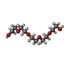
| #1: Antibody | Mass: 24072.021 Da / Num. of mol.: 2 Source method: isolated from a genetically manipulated source Source: (gene. exp.)   Homo sapiens (human) Homo sapiens (human)#2: Antibody | Mass: 22777.102 Da / Num. of mol.: 2 Source method: isolated from a genetically manipulated source Source: (gene. exp.)   Homo sapiens (human) Homo sapiens (human)#3: Chemical | ChemComp-SO4 / #4: Chemical | ChemComp-1PE / | #5: Water | ChemComp-HOH / | Has ligand of interest | N | Has protein modification | Y | |
|---|
-Experimental details
-Experiment
| Experiment | Method:  X-RAY DIFFRACTION / Number of used crystals: 1 X-RAY DIFFRACTION / Number of used crystals: 1 |
|---|
- Sample preparation
| Crystal | Density Matthews: 2.41 Å3/Da / Density % sol: 48.95 % |
|---|---|
| Crystal grow | Temperature: 298 K / Method: vapor diffusion, sitting drop / pH: 4.5 Details: 0.1M sodium acetate, 0.2M lithium sulfate, 30% PEG8000 |
-Data collection
| Diffraction | Mean temperature: 100 K / Serial crystal experiment: N |
|---|---|
| Diffraction source | Source:  SYNCHROTRON / Site: SYNCHROTRON / Site:  APS APS  / Beamline: 23-ID-D / Wavelength: 1.03315 Å / Beamline: 23-ID-D / Wavelength: 1.03315 Å |
| Detector | Type: DECTRIS PILATUS 6M / Detector: PIXEL / Date: Oct 15, 2016 |
| Radiation | Protocol: SINGLE WAVELENGTH / Monochromatic (M) / Laue (L): M / Scattering type: x-ray |
| Radiation wavelength | Wavelength: 1.03315 Å / Relative weight: 1 |
| Reflection | Resolution: 2.2→45.9 Å / Num. obs: 44732 / % possible obs: 98.7 % / Observed criterion σ(F): 0 / Observed criterion σ(I): -3 / Redundancy: 6.4 % / Biso Wilson estimate: 38.17 Å2 / CC1/2: 0.86 / Rmerge(I) obs: 0.185 / Rpim(I) all: 0.078 / Rrim(I) all: 0.201 / Net I/σ(I): 9 |
| Reflection shell | Resolution: 2.2→2.24 Å / Redundancy: 5.2 % / Rmerge(I) obs: 1.2 / Mean I/σ(I) obs: 1 / Num. unique obs: 2195 / CC1/2: 0.56 / Rpim(I) all: 0.55 / Rrim(I) all: 1.3 / % possible all: 96.5 |
- Processing
| Software |
| |||||||||||||||||||||||||||||||||||||||||||||||||||||||||||||||||||||||||||||||||||||||||||||||||||||||||||||||||||||||
|---|---|---|---|---|---|---|---|---|---|---|---|---|---|---|---|---|---|---|---|---|---|---|---|---|---|---|---|---|---|---|---|---|---|---|---|---|---|---|---|---|---|---|---|---|---|---|---|---|---|---|---|---|---|---|---|---|---|---|---|---|---|---|---|---|---|---|---|---|---|---|---|---|---|---|---|---|---|---|---|---|---|---|---|---|---|---|---|---|---|---|---|---|---|---|---|---|---|---|---|---|---|---|---|---|---|---|---|---|---|---|---|---|---|---|---|---|---|---|---|---|
| Refinement | Method to determine structure:  MOLECULAR REPLACEMENT MOLECULAR REPLACEMENTStarting model: homology model Resolution: 2.2→45.9 Å / SU ML: 0.323 / Cross valid method: FREE R-VALUE / σ(F): 1.34 / Phase error: 29.2954 Stereochemistry target values: GeoStd + Monomer Library + CDL v1.2
| |||||||||||||||||||||||||||||||||||||||||||||||||||||||||||||||||||||||||||||||||||||||||||||||||||||||||||||||||||||||
| Solvent computation | Shrinkage radii: 0.9 Å / VDW probe radii: 1.11 Å / Solvent model: FLAT BULK SOLVENT MODEL | |||||||||||||||||||||||||||||||||||||||||||||||||||||||||||||||||||||||||||||||||||||||||||||||||||||||||||||||||||||||
| Displacement parameters | Biso mean: 41.27 Å2 | |||||||||||||||||||||||||||||||||||||||||||||||||||||||||||||||||||||||||||||||||||||||||||||||||||||||||||||||||||||||
| Refinement step | Cycle: LAST / Resolution: 2.2→45.9 Å
| |||||||||||||||||||||||||||||||||||||||||||||||||||||||||||||||||||||||||||||||||||||||||||||||||||||||||||||||||||||||
| Refine LS restraints |
| |||||||||||||||||||||||||||||||||||||||||||||||||||||||||||||||||||||||||||||||||||||||||||||||||||||||||||||||||||||||
| LS refinement shell |
|
 Movie
Movie Controller


 PDBj
PDBj


