 Movie
Movie Controller


+ Open data

- Basic information
| Entry | Database: PDB / ID: 7r7o | ||||||||||||
|---|---|---|---|---|---|---|---|---|---|---|---|---|---|
| Title | Structure of methyltransferase domain of Spb1 boudn to SAM | ||||||||||||
 Components Components | AdoMet-dependent rRNA methyltransferase SPB1 | ||||||||||||
 Keywords Keywords | TRANSFERASE / ribosome biogenesis / methyltransferase / nucleolus | ||||||||||||
| Function / homology | S-ADENOSYLMETHIONINE / :  Function and homology information Function and homology information | ||||||||||||
| Biological species |  | ||||||||||||
| Method |  X-RAY DIFFRACTION / SYNCHROTRON / MOLECULAR REPLACEMENT / Resolution: 1.58 Å X-RAY DIFFRACTION / SYNCHROTRON / MOLECULAR REPLACEMENT / Resolution: 1.58 Å | ||||||||||||
 Authors Authors | Cruz, V.E. / Sekulski, K. / Peddada, N. / Erzberger, J.P. | ||||||||||||
| Funding support |  United States, 3items United States, 3items
| ||||||||||||
 Citation Citation | Journal: To Be Published Title: Post-catalytic rRNA binding by the DEAD-box ATPase Spb4 and methyltransferase Spb1 guide the late nucleolar assembly of 60S ribosomes Authors: Cruz, V.E. / Sekulski, K. / Peddada, N. / Sailer, C. / Balasubramanian, S. / Weirich, C.S. / Stengel, F. / Erzberger, J.P. | ||||||||||||
| History |
|
- Structure visualization
| Structure viewer | Molecule: MolmilJmol/JSmol |
|---|
- Downloads & links
-Download
| PDBx/mmCIF format | 7r7o.cif.gz | 122.7 KB | Display | PDBx/mmCIF format |
|---|---|---|---|---|
| PDB format | pdb7r7o.ent.gz | 84 KB | Display | PDB format |
| PDBx/mmJSON format | 7r7o.json.gz | Tree view | PDBx/mmJSON format | |
| Others |  Other downloads Other downloads |
-Validation report
| Arichive directory | https://data.pdbj.org/pub/pdb/validation_reports/r7/7r7oftp://data.pdbj.org/pub/pdb/validation_reports/r7/7r7o | HTTPS FTP |
|---|
-Related structure data
| Related structure data |  6elzS S: Starting model for refinement |
|---|---|
| Similar structure data |


-Links
 PDBj
PDBj
- Assembly
| Deposited unit | 
| ||||||||||||
|---|---|---|---|---|---|---|---|---|---|---|---|---|---|
| 1 | 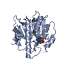
| ||||||||||||
| 2 | 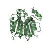
| ||||||||||||
| Unit cell |
|
-Components
| #1: Protein | Mass: 24990.193 Da / Num. of mol.: 2 Source method: isolated from a genetically manipulated source Source: (gene. exp.) Gene: SPB1, PACBIOSEQ_LOCUS534 / Production host:  References: UniProt: A0A7I9D0Y8, Transferases; Transferring one-carbon groups; Methyltransferases #2: Chemical |   Mass: 398.437 Da / Num. of mol.: 2 / Source method: obtained synthetically / Formula: C15H22N6O5S / Feature type: SUBJECT OF INVESTIGATION Mass: 398.437 Da / Num. of mol.: 2 / Source method: obtained synthetically / Formula: C15H22N6O5S / Feature type: SUBJECT OF INVESTIGATION#3: Water | ChemComp-HOH / |  Mass: 18.015 Da / Num. of mol.: 541 / Source method: isolated from a natural source / Formula: H2O Mass: 18.015 Da / Num. of mol.: 541 / Source method: isolated from a natural source / Formula: H2OHas ligand of interest | Y | |
|---|
-Experimental details
-Experiment
| Experiment | Method: X-RAY DIFFRACTION / Number of used crystals: 1 |
|---|
- Sample preparation
| Crystal | Density Matthews: 2.46 Å3/Da / Density % sol: 50 % |
|---|---|
| Crystal grow | Temperature: 293 K / Method: vapor diffusion, hanging drop / pH: 8 Details: 20 mM Hepes KOH pH 8 150 mM KCl 0.5 mM TCEP 5% Glycerol 15% PEG 3350 |
-Data collection
| Diffraction | Mean temperature: 100 K / Serial crystal experiment: N |
|---|---|
| Diffraction source | Source: SYNCHROTRON / Site: APS / Beamline: 19-ID / Wavelength: 1 Å |
| Detector | Type: ADSC QUANTUM 315r / Detector: CCD / Date: May 26, 2019 |
| Radiation | Protocol: SINGLE WAVELENGTH / Monochromatic (M) / Laue (L): M / Scattering type: x-ray |
| Radiation wavelength | Wavelength: 1 Å / Relative weight: 1 |
| Reflection | Resolution: 1.58→50 Å / Num. obs: 66501 / % possible obs: 83.08 % / Redundancy: 8.1 % / Biso Wilson estimate: 14.3 Å2 / CC1/2: 0.982 / CC star: 0.995 / Rpim(I) all: 0.037 / Rrim(I) all: 0.109 / Χ2: 0.89 / Net I/σ(I): 18.1 |
| Reflection shell | Resolution: 1.58→1.61 Å / Mean I/σ(I) obs: 0.93 / Num. unique obs: 3158 / CC1/2: 0.497 / CC star: 0.815 / Rpim(I) all: 0.037 / Χ2: 0.89 |
- Processing
| Software |
| |||||||||||||||||||||||||||||||||||||||||||||||||||||||||||||||||||||||||||||||||||||||||||||||||||||||||
|---|---|---|---|---|---|---|---|---|---|---|---|---|---|---|---|---|---|---|---|---|---|---|---|---|---|---|---|---|---|---|---|---|---|---|---|---|---|---|---|---|---|---|---|---|---|---|---|---|---|---|---|---|---|---|---|---|---|---|---|---|---|---|---|---|---|---|---|---|---|---|---|---|---|---|---|---|---|---|---|---|---|---|---|---|---|---|---|---|---|---|---|---|---|---|---|---|---|---|---|---|---|---|---|---|---|---|
| Refinement | Method to determine structure: MOLECULAR REPLACEMENT Starting model: 6ELZ Resolution: 1.58→42.42 Å / SU ML: 0.1906 / Cross valid method: FREE R-VALUE / σ(F): 1.34 / Phase error: 27.0965 Stereochemistry target values: GeoStd + Monomer Library + CDL v1.2
| |||||||||||||||||||||||||||||||||||||||||||||||||||||||||||||||||||||||||||||||||||||||||||||||||||||||||
| Solvent computation | Shrinkage radii: 0.9 Å / VDW probe radii: 1.11 Å / Solvent model: FLAT BULK SOLVENT MODEL | |||||||||||||||||||||||||||||||||||||||||||||||||||||||||||||||||||||||||||||||||||||||||||||||||||||||||
| Refinement step | Cycle: LAST / Resolution: 1.58→42.42 Å
| |||||||||||||||||||||||||||||||||||||||||||||||||||||||||||||||||||||||||||||||||||||||||||||||||||||||||
| Refine LS restraints |
| |||||||||||||||||||||||||||||||||||||||||||||||||||||||||||||||||||||||||||||||||||||||||||||||||||||||||
| LS refinement shell |
|
