Entry Database : PDB / ID : 7r2wTitle Mutant S-adenosylmethionine synthetase from E.coli complexed with AMPPNP and methionine S-adenosylmethionine synthase Keywords / / / / Function / homology Function Domain/homology Component
/ / / / / / / / / / / / / / / / / / / / / / / / / / / / / Biological species Escherichia coli (E. coli)Method / / / Resolution : 1.6 Å Authors Shahar, A. / Kleiner, D. / Bershtein, S. / Zarivach, R. Funding support Organization Grant number Country United States - Israel Binational Science Foundation (BSF) 2020640
Journal : Protein Sci. / Year : 2022Title : Evolution of homo-oligomerization of methionine S-adenosyltransferases is replete with structure-function constrains.Authors : Kleiner, D. / Shapiro Tuchman, Z. / Shmulevich, F. / Shahar, A. / Zarivach, R. / Kosloff, M. / Bershtein, S. History Deposition Feb 6, 2022 Deposition site / Processing site Revision 1.0 Jul 13, 2022 Provider / Type Revision 1.1 Jan 31, 2024 Group / Refinement descriptionCategory / chem_comp_bond / pdbx_initial_refinement_model
Show all Show less
 Movie
Movie Controller

 Yorodumi
 Open data
 Basic information
 Components
 Keywords
 Function and homology information
 X-RAY DIFFRACTION /
  Authors
 United States, 1items
  Citation
 Structure visualization
 Downloads & links
 Other downloads
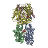

 PDBj
PDBj


 Assembly
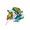


 Mass: 506.196 Da / Num. of mol.: 1 / Source method: obtained synthetically / Formula: C10H17N6O12P3 / Feature type: SUBJECT OF INVESTIGATION / Comment: AMP-PNP, energy-carrying molecule analogue*YM
 Mass: 24.305 Da / Num. of mol.: 2 / Source method: obtained synthetically / Formula: Mg / Feature type: SUBJECT OF INVESTIGATION
 Mass: 39.098 Da / Num. of mol.: 1 / Source method: obtained synthetically / Formula: K / Feature type: SUBJECT OF INVESTIGATION
 Mass: 18.015 Da / Num. of mol.: 185 / Source method: isolated from a natural source / Formula: H2O
 Sample preparation
 / Beamline: I03 / Wavelength: 0.97626 Å
 Processing