 Movie
Movie Controller


+ Open data

- Basic information
| Entry | Database: PDB / ID: 7qzq | ||||||
|---|---|---|---|---|---|---|---|
| Title | Crystal structure of the kelch domain of human KBTBD12 | ||||||
 Components Components | Kelch repeat and BTB domain-containing protein 12 | ||||||
 Keywords Keywords | LIGASE / E3 ligase / Kelch / BTB-Kelch | ||||||
| Function / homology |  Function and homology information Function and homology informationKelch-type beta propeller / BTB-kelch protein / BTB/Kelch-associated / BTB And C-terminal Kelch / BTB And C-terminal Kelch / Kelch / Kelch motif / Kelch repeat type 1 / Kelch-type beta propeller / 6 Propeller ...Kelch-type beta propeller / BTB-kelch protein / BTB/Kelch-associated / BTB And C-terminal Kelch / BTB And C-terminal Kelch / Kelch / Kelch motif / Kelch repeat type 1 / Kelch-type beta propeller / 6 Propeller / Neuraminidase / BTB/POZ domain / BTB domain profile. / Broad-Complex, Tramtrack and Bric a brac / BTB/POZ domain / SKP1/BTB/POZ domain superfamily / Mainly Beta Similarity search - Domain/homology | ||||||
| Biological species |  Homo sapiens (human) Homo sapiens (human) | ||||||
| Method |  X-RAY DIFFRACTION / SYNCHROTRON / MOLECULAR REPLACEMENT / molecular replacement / Resolution: 1.88 Å X-RAY DIFFRACTION / SYNCHROTRON / MOLECULAR REPLACEMENT / molecular replacement / Resolution: 1.88 Å | ||||||
 Authors Authors | Manning, C.E. / Chen, Z. / Chen, X. / Bradshaw, W.J. / Bakshi, S. / Mckinley, G. / Chalk, R. / Burgess-Brown, N. / von Delft, F. / Bullock, A.N. | ||||||
| Funding support |  United Kingdom, 1items United Kingdom, 1items
| ||||||
 Citation Citation | Journal: To Be Published Title: Crystal structure of the kelch domain of human KBTBD12 Authors: Manning, C.E. / Chen, Z. / Chen, X. / Bakshi, S. / Mckinley, G. / Chalk, R. / Burgess-Brown, N. / von Delft, F. / Bullock, A.N. | ||||||
| History |
|
- Structure visualization
| Structure viewer | Molecule: MolmilJmol/JSmol |
|---|
- Downloads & links
-Download
| PDBx/mmCIF format | 7qzq.cif.gz | 157.4 KB | Display | PDBx/mmCIF format |
|---|---|---|---|---|
| PDB format | pdb7qzq.ent.gz | 119.5 KB | Display | PDB format |
| PDBx/mmJSON format | 7qzq.json.gz | Tree view | PDBx/mmJSON format | |
| Others |  Other downloads Other downloads |
-Validation report
| Arichive directory | https://data.pdbj.org/pub/pdb/validation_reports/qz/7qzqftp://data.pdbj.org/pub/pdb/validation_reports/qz/7qzq | HTTPS FTP |
|---|
-Related structure data
| Related structure data |  2vpjS S: Starting model for refinement |
|---|---|
| Similar structure data |
-Links
 PDBj
PDBj


- Assembly
| Deposited unit | 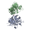
| ||||||||
|---|---|---|---|---|---|---|---|---|---|
| 1 | 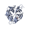
| ||||||||
| 2 | 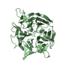
| ||||||||
| Unit cell |
|
-Components
| #1: Protein | Mass: 40863.207 Da / Num. of mol.: 2 Source method: isolated from a genetically manipulated source Source: (gene. exp.) Homo sapiens (human) / Gene: KBTBD12, KLHDC6 / Cell line (production host): Sf9Production host:  Spodoptera frugiperda multiple nucleopolyhedrovirus Spodoptera frugiperda multiple nucleopolyhedrovirusReferences: UniProt: Q3ZCT8 #2: Chemical | ChemComp-EDO / |   Mass: 62.068 Da / Num. of mol.: 1 / Source method: obtained synthetically / Formula: C2H6O2 Mass: 62.068 Da / Num. of mol.: 1 / Source method: obtained synthetically / Formula: C2H6O2#3: Chemical | ChemComp-NA / |   Mass: 22.990 Da / Num. of mol.: 1 / Source method: obtained synthetically / Formula: Na Mass: 22.990 Da / Num. of mol.: 1 / Source method: obtained synthetically / Formula: Na#4: Water | ChemComp-HOH / |  Mass: 18.015 Da / Num. of mol.: 446 / Source method: isolated from a natural source / Formula: H2O Mass: 18.015 Da / Num. of mol.: 446 / Source method: isolated from a natural source / Formula: H2OHas ligand of interest | N | |
|---|
-Experimental details
-Experiment
| Experiment | Method: X-RAY DIFFRACTION / Number of used crystals: 1 |
|---|
- Sample preparation
| Crystal | Density Matthews: 2.2 Å3/Da / Density % sol: 44.06 % |
|---|---|
| Crystal grow | Temperature: 293.15 K / Method: vapor diffusion, sitting drop / pH: 4.5 / Details: 8% PEG4000 -- 0.1M acetate pH 4.5 |
-Data collection
| Diffraction | Mean temperature: 100 K / Serial crystal experiment: N |
|---|---|
| Diffraction source | Source: SYNCHROTRON / Site: Diamond / Beamline: I03 / Wavelength: 0.9763 Å |
| Detector | Type: DECTRIS PILATUS 6M / Detector: PIXEL / Date: Aug 23, 2021 |
| Radiation | Protocol: SINGLE WAVELENGTH / Monochromatic (M) / Laue (L): M / Scattering type: x-ray |
| Radiation wavelength | Wavelength: 0.9763 Å / Relative weight: 1 |
| Reflection | Resolution: 1.88→65.63 Å / Num. obs: 57704 / % possible obs: 99.9 % / Redundancy: 6.4 % / Biso Wilson estimate: 32.03 Å2 / CC1/2: 0.997 / Rmerge(I) obs: 0.156 / Rrim(I) all: 0.17 / Net I/σ(I): 7.3 |
| Reflection shell | Resolution: 1.88→1.92 Å / Redundancy: 5.6 % / Rmerge(I) obs: 3.1 / Mean I/σ(I) obs: 0.5 / Num. unique obs: 2827 / CC1/2: 0.268 / % possible all: 97.7 |
-Phasing
| Phasing | Method: molecular replacement | |||||||||
|---|---|---|---|---|---|---|---|---|---|---|
| Phasing MR |
|
- Processing
| Software |
| |||||||||||||||||||||||||||||||||||||||||||||||||||||||||||||||||||||||||||||||||||||||||||||||||||||||||||||||||||||||||||||||||||||||||||||||||||
|---|---|---|---|---|---|---|---|---|---|---|---|---|---|---|---|---|---|---|---|---|---|---|---|---|---|---|---|---|---|---|---|---|---|---|---|---|---|---|---|---|---|---|---|---|---|---|---|---|---|---|---|---|---|---|---|---|---|---|---|---|---|---|---|---|---|---|---|---|---|---|---|---|---|---|---|---|---|---|---|---|---|---|---|---|---|---|---|---|---|---|---|---|---|---|---|---|---|---|---|---|---|---|---|---|---|---|---|---|---|---|---|---|---|---|---|---|---|---|---|---|---|---|---|---|---|---|---|---|---|---|---|---|---|---|---|---|---|---|---|---|---|---|---|---|---|---|---|---|
| Refinement | Method to determine structure: MOLECULAR REPLACEMENT Starting model: 2VPJ Resolution: 1.88→45.08 Å / SU ML: 0.28 / Cross valid method: THROUGHOUT / σ(F): 1.34 / Phase error: 31.15 / Stereochemistry target values: ML
| |||||||||||||||||||||||||||||||||||||||||||||||||||||||||||||||||||||||||||||||||||||||||||||||||||||||||||||||||||||||||||||||||||||||||||||||||||
| Solvent computation | Shrinkage radii: 0.9 Å / VDW probe radii: 1.11 Å / Solvent model: FLAT BULK SOLVENT MODEL | |||||||||||||||||||||||||||||||||||||||||||||||||||||||||||||||||||||||||||||||||||||||||||||||||||||||||||||||||||||||||||||||||||||||||||||||||||
| Displacement parameters | Biso max: 121.14 Å2 / Biso mean: 40.4986 Å2 / Biso min: 22.73 Å2 | |||||||||||||||||||||||||||||||||||||||||||||||||||||||||||||||||||||||||||||||||||||||||||||||||||||||||||||||||||||||||||||||||||||||||||||||||||
| Refinement step | Cycle: final / Resolution: 1.88→45.08 Å
| |||||||||||||||||||||||||||||||||||||||||||||||||||||||||||||||||||||||||||||||||||||||||||||||||||||||||||||||||||||||||||||||||||||||||||||||||||
| LS refinement shell | Refine-ID: X-RAY DIFFRACTION / Rfactor Rfree error: 0 / Total num. of bins used: 20
|
