 Movie
Movie Controller


+ Open data

- Basic information
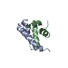
| Entry | Database: PDB / ID: 7qy5 | ||||||
|---|---|---|---|---|---|---|---|
| Title | Crystal structure of the S.pombe Ars2-Red1 complex. | ||||||
 Components Components |
| ||||||
 Keywords Keywords | RNA / RNA degradation / RNA targeting complex | ||||||
| Function / homology |  Function and homology information Function and homology informationMTREC complex / nuclear polyadenylation-dependent CUT catabolic process / regulatory ncRNA-mediated heterochromatin formation / regulatory ncRNA-mediated gene silencing / lncRNA binding / mRNA processing / nuclear body / chromatin / metal ion binding / nucleus Similarity search - Function | ||||||
| Biological species |  | ||||||
| Method |  X-RAY DIFFRACTION / SYNCHROTRON / MOLECULAR REPLACEMENT / Resolution: 2.77 Å X-RAY DIFFRACTION / SYNCHROTRON / MOLECULAR REPLACEMENT / Resolution: 2.77 Å | ||||||
 Authors Authors | Foucher, A.E. / Kadlec, J. | ||||||
| Funding support |  France, 1items France, 1items
| ||||||
 Citation Citation | Journal: Nat Commun / Year: 2022 Title: Structural analysis of Red1 as a conserved scaffold of the RNA-targeting MTREC/PAXT complex. Authors: Foucher, A.E. / Touat-Todeschini, L. / Juarez-Martinez, A.B. / Rakitch, A. / Laroussi, H. / Karczewski, C. / Acajjaoui, S. / Soler-Lopez, M. / Cusack, S. / Mackereth, C.D. / Verdel, A. / Kadlec, J. | ||||||
| History |
|
- Structure visualization
| Structure viewer | Molecule: MolmilJmol/JSmol |
|---|
- Downloads & links
-Download
| PDBx/mmCIF format | 7qy5.cif.gz | 189.7 KB | Display | PDBx/mmCIF format |
|---|---|---|---|---|
| PDB format | pdb7qy5.ent.gz | 149.2 KB | Display | PDB format |
| PDBx/mmJSON format | 7qy5.json.gz | Tree view | PDBx/mmJSON format | |
| Others |  Other downloads Other downloads |
-Validation report
| Arichive directory | https://data.pdbj.org/pub/pdb/validation_reports/qy/7qy5ftp://data.pdbj.org/pub/pdb/validation_reports/qy/7qy5 | HTTPS FTP |
|---|
-Related structure data
-Links
 PDBj
PDBj


- Assembly
| Deposited unit | 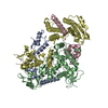
| ||||||||
|---|---|---|---|---|---|---|---|---|---|
| 1 |
| ||||||||
| Unit cell |
|
-Components
| #1: Protein | Mass: 14315.451 Da / Num. of mol.: 2 Source method: isolated from a genetically manipulated source Source: (gene. exp.) Strain: 972 / ATCC 24843 / Gene: pir2, ars2, SPBC725.08 / Production host:  #2: Protein/peptide | Mass: 2401.233 Da / Num. of mol.: 2 / Source method: obtained synthetically / Source: (synth.) #3: Protein | Mass: 38815.859 Da / Num. of mol.: 2 Source method: isolated from a genetically manipulated source Source: (gene. exp.) Strain: 972 / ATCC 24843 / Gene: pir2, ars2, SPBC725.08 / Production host: #4: Chemical |   Mass: 65.409 Da / Num. of mol.: 2 / Source method: obtained synthetically / Formula: Zn Mass: 65.409 Da / Num. of mol.: 2 / Source method: obtained synthetically / Formula: ZnHas ligand of interest | N | |
|---|
-Experimental details
-Experiment
| Experiment | Method: X-RAY DIFFRACTION / Number of used crystals: 1 |
|---|
- Sample preparation
| Crystal | Density Matthews: 3.46 Å3/Da / Density % sol: 64.41 % |
|---|---|
| Crystal grow | Temperature: 278.15 K / Method: vapor diffusion, sitting drop / pH: 8 / Details: 200mM Ammonium Formate, 20% PEG3350 |
-Data collection
| Diffraction | Mean temperature: 100 K / Serial crystal experiment: N |
|---|---|
| Diffraction source | Source: SYNCHROTRON / Site: ESRF / Beamline: ID30B / Wavelength: 0.9677 Å |
| Detector | Type: DECTRIS EIGER R 4M / Detector: PIXEL / Date: Nov 7, 2021 |
| Radiation | Protocol: SINGLE WAVELENGTH / Monochromatic (M) / Laue (L): M / Scattering type: x-ray |
| Radiation wavelength | Wavelength: 0.9677 Å / Relative weight: 1 |
| Reflection | Resolution: 2.77→84.85 Å / Num. obs: 26786 / % possible obs: 76 % / Redundancy: 5.56 % / CC1/2: 0.98 / Rmerge(I) obs: 0.29 / Net I/σ(I): 5.3 |
| Reflection shell | Resolution: 2.771→2.994 Å / Redundancy: 6.2 % / Mean I/σ(I) obs: 1.5 / Num. unique obs: 1339 / CC1/2: 0.55 / % possible all: 18.4 |
- Processing
| Software |
| ||||||||||||||||||||||||||||||||||||||||||||||||||||||||||||
|---|---|---|---|---|---|---|---|---|---|---|---|---|---|---|---|---|---|---|---|---|---|---|---|---|---|---|---|---|---|---|---|---|---|---|---|---|---|---|---|---|---|---|---|---|---|---|---|---|---|---|---|---|---|---|---|---|---|---|---|---|---|
| Refinement | Method to determine structure: MOLECULAR REPLACEMENT Starting model: alpafold Resolution: 2.77→84.85 Å / Cor.coef. Fo:Fc: 0.927 / Cor.coef. Fo:Fc free: 0.865 / SU B: 21.803 / SU ML: 0.401 / Cross valid method: THROUGHOUT / σ(F): 0 / ESU R Free: 0.488 / Stereochemistry target values: MAXIMUM LIKELIHOOD Details: HYDROGENS HAVE BEEN ADDED IN THE RIDING POSITIONS U VALUES : REFINED INDIVIDUALLY
| ||||||||||||||||||||||||||||||||||||||||||||||||||||||||||||
| Solvent computation | Ion probe radii: 0.8 Å / Shrinkage radii: 0.8 Å / VDW probe radii: 1.2 Å / Solvent model: MASK | ||||||||||||||||||||||||||||||||||||||||||||||||||||||||||||
| Displacement parameters | Biso max: 160.83 Å2 / Biso mean: 52.291 Å2 / Biso min: 24 Å2
| ||||||||||||||||||||||||||||||||||||||||||||||||||||||||||||
| Refinement step | Cycle: final / Resolution: 2.77→84.85 Å
| ||||||||||||||||||||||||||||||||||||||||||||||||||||||||||||
| Refine LS restraints |
| ||||||||||||||||||||||||||||||||||||||||||||||||||||||||||||
| LS refinement shell | Resolution: 2.771→2.843 Å / Rfactor Rfree error: 0 / Total num. of bins used: 20
|
