regulation of cell budding / regulation of protein localization to cell division site involved in cytokinesis / cellular bud neck septin collar / mating projection base / regulation of cellular response to glucose starvation / positive regulation of mitotic actomyosin contractile ring assembly / Resolution of Sister Chromatid Cohesion / termination of RNA polymerase II transcription, poly(A)-coupled / chitin biosynthetic process / regulation of glycogen biosynthetic process ...regulation of cell budding / regulation of protein localization to cell division site involved in cytokinesis / cellular bud neck septin collar / mating projection base / regulation of cellular response to glucose starvation / positive regulation of mitotic actomyosin contractile ring assembly / Resolution of Sister Chromatid Cohesion / termination of RNA polymerase II transcription, poly(A)-coupled / chitin biosynthetic process / regulation of glycogen biosynthetic process / cellular bud site selection / positive regulation of clathrin-dependent endocytosis / ascospore formation / positive regulation of exit from mitosis / protein phosphatase type 1 complex / incipient cellular bud site / negative regulation of actin filament polymerization / intracellular monoatomic ion homeostasis / mRNA cleavage and polyadenylation specificity factor complex / DNA replication checkpoint signaling / cellular bud neck / protein localization to kinetochore / spindle pole body / glycogen metabolic process / cell division site / protein-serine/threonine phosphatase / mitotic spindle assembly checkpoint signaling / protein serine/threonine phosphatase activity / replication fork processing / response to unfolded protein / chromosome organization / regulation of mitotic cell cycle / telomere maintenance / DNA damage checkpoint signaling / meiotic cell cycle / chromosome segregation / kinetochore / mRNA processing / regulation of cell shape / response to heat / regulation of cell cycle / nucleolus / positive regulation of transcription by RNA polymerase II / metal ion binding / nucleus / cytoplasm Similarity search - Function
: / Serine-threonine protein phosphatase, N-terminal / Serine-threonine protein phosphatase N-terminal domain / Serine/threonine specific protein phosphatases signature. / Protein phosphatase 2A homologues, catalytic domain. / Serine/threonine-specific protein phosphatase/bis(5-nucleosyl)-tetraphosphatase / Metallo-dependent phosphatases / Purple Acid Phosphatase; chain A, domain 2 / Calcineurin-like phosphoesterase domain, ApaH type / Calcineurin-like phosphoesterase ...: / Serine-threonine protein phosphatase, N-terminal / Serine-threonine protein phosphatase N-terminal domain / Serine/threonine specific protein phosphatases signature. / Protein phosphatase 2A homologues, catalytic domain. / Serine/threonine-specific protein phosphatase/bis(5-nucleosyl)-tetraphosphatase / Metallo-dependent phosphatases / Purple Acid Phosphatase; chain A, domain 2 / Calcineurin-like phosphoesterase domain, ApaH type / Calcineurin-like phosphoesterase / Metallo-dependent phosphatase-like / 4-Layer Sandwich / Alpha Beta Similarity search - Domain/homology
In the structure databanks used in Yorodumi, some data are registered as the other names, "COVID-19 virus" and "2019-nCoV". Here are the details of the virus and the list of structure data.
Jan 31, 2019. EMDB accession codes are about to change! (news from PDBe EMDB page)
EMDB accession codes are about to change! (news from PDBe EMDB page)
The allocation of 4 digits for EMDB accession codes will soon come to an end. Whilst these codes will remain in use, new EMDB accession codes will include an additional digit and will expand incrementally as the available range of codes is exhausted. The current 4-digit format prefixed with “EMD-” (i.e. EMD-XXXX) will advance to a 5-digit format (i.e. EMD-XXXXX), and so on. It is currently estimated that the 4-digit codes will be depleted around Spring 2019, at which point the 5-digit format will come into force.
The EM Navigator/Yorodumi systems omit the EMD- prefix.
Related info.:Q: What is EMD? / ID/Accession-code notation in Yorodumi/EM Navigator
Yorodumi is a browser for structure data from EMDB, PDB, SASBDB, etc.
This page is also the successor to EM Navigator detail page, and also detail information page/front-end page for Omokage search.
The word "yorodu" (or yorozu) is an old Japanese word meaning "ten thousand". "mi" (miru) is to see.
Related info.:EMDB / PDB / SASBDB / Comparison of 3 databanks / Yorodumi Search / Aug 31, 2016. New EM Navigator & Yorodumi / Yorodumi Papers / Jmol/JSmol / Function and homology information / Changes in new EM Navigator and Yorodumi
 Movie
Movie Controller


 Open data
 Basic information
 Components
 Keywords
 Function and homology information
 X-RAY DIFFRACTION /
  Authors
 United Kingdom, 1items
  Citation
 Structure visualization
 Downloads & links
 Other downloads
 PDBj
PDBj
 Assembly
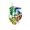


 Mass: 54.938 Da / Num. of mol.: 2 / Source method: obtained synthetically / Formula: Mn / Feature type: SUBJECT OF INVESTIGATION
 Mass: 94.971 Da / Num. of mol.: 1 / Source method: obtained synthetically / Formula: PO4 / Feature type: SUBJECT OF INVESTIGATION
 Mass: 18.015 Da / Num. of mol.: 84 / Source method: isolated from a natural source / Formula: H2O
 Sample preparation
 Processing