 Movie
Movie Controller

[English] 日本語
 Yorodumi
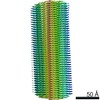
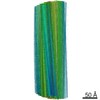
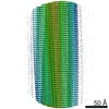
- PDB-7qwg: TMEM106B filaments with Fold IIa from Multiple system atrophy (ca... -
+ Open data

- Basic information
| Entry | Database: PDB / ID: 7qwg | ||||||
|---|---|---|---|---|---|---|---|
| Title | TMEM106B filaments with Fold IIa from Multiple system atrophy (case 19) | ||||||
 Components Components | Transmembrane protein 106B | ||||||
 Keywords Keywords | PROTEIN FIBRIL / Amyloid / neurodegeneration / filament / TMEM106B | ||||||
| Function / homology |  Function and homology information Function and homology informationlysosomal protein catabolic process / lysosomal lumen acidification / lysosome localization / regulation of lysosome organization / dendrite morphogenesis / positive regulation of dendrite development / lysosomal transport / lysosome organization / neuron cellular homeostasis / late endosome membrane ...lysosomal protein catabolic process / lysosomal lumen acidification / lysosome localization / regulation of lysosome organization / dendrite morphogenesis / positive regulation of dendrite development / lysosomal transport / lysosome organization / neuron cellular homeostasis / late endosome membrane / ATPase binding / lysosome / endosome / lysosomal membrane / plasma membrane Similarity search - Function | ||||||
| Biological species |  Homo sapiens (human) Homo sapiens (human) | ||||||
| Method | ELECTRON MICROSCOPY / helical reconstruction / cryo EM / Resolution: 3.38 Å | ||||||
 Authors Authors | Lovestam, S. / Schweighauser, M. / Scheres, S.H.W. | ||||||
| Funding support |  United Kingdom, 1items United Kingdom, 1items
| ||||||
 Citation Citation | Journal: Nature / Year: 2022 Title: Age-dependent formation of TMEM106B amyloid filaments in human brains. Authors: Manuel Schweighauser / Diana Arseni / Mehtap Bacioglu / Melissa Huang / Sofia Lövestam / Yang Shi / Yang Yang / Wenjuan Zhang / Abhay Kotecha / Holly J Garringer / Ruben Vidal / Grace I ...Authors: Manuel Schweighauser / Diana Arseni / Mehtap Bacioglu / Melissa Huang / Sofia Lövestam / Yang Shi / Yang Yang / Wenjuan Zhang / Abhay Kotecha / Holly J Garringer / Ruben Vidal / Grace I Hallinan / Kathy L Newell / Airi Tarutani / Shigeo Murayama / Masayuki Miyazaki / Yuko Saito / Mari Yoshida / Kazuko Hasegawa / Tammaryn Lashley / Tamas Revesz / Gabor G Kovacs / John van Swieten / Masaki Takao / Masato Hasegawa / Bernardino Ghetti / Maria Grazia Spillantini / Benjamin Ryskeldi-Falcon / Alexey G Murzin / Michel Goedert / Sjors H W Scheres /      Abstract: Many age-dependent neurodegenerative diseases, such as Alzheimer's and Parkinson's, are characterized by abundant inclusions of amyloid filaments. Filamentous inclusions of the proteins tau, amyloid- ...Many age-dependent neurodegenerative diseases, such as Alzheimer's and Parkinson's, are characterized by abundant inclusions of amyloid filaments. Filamentous inclusions of the proteins tau, amyloid-β, α-synuclein and transactive response DNA-binding protein (TARDBP; also known as TDP-43) are the most common. Here we used structure determination by cryogenic electron microscopy to show that residues 120-254 of the lysosomal type II transmembrane protein 106B (TMEM106B) also form amyloid filaments in human brains. We determined the structures of TMEM106B filaments from a number of brain regions of 22 individuals with abundant amyloid deposits, including those resulting from sporadic and inherited tauopathies, amyloid-β amyloidoses, synucleinopathies and TDP-43 proteinopathies, as well as from the frontal cortex of 3 individuals with normal neurology and no or only a few amyloid deposits. We observed three TMEM106B folds, with no clear relationships between folds and diseases. TMEM106B filaments correlated with the presence of a 29-kDa sarkosyl-insoluble fragment and globular cytoplasmic inclusions, as detected by an antibody specific to the carboxy-terminal region of TMEM106B. The identification of TMEM106B filaments in the brains of older, but not younger, individuals with normal neurology indicates that they form in an age-dependent manner. | ||||||
| History |
|
- Structure visualization
| Movie |
Movie viewer |
|---|---|
| Structure viewer | Molecule: MolmilJmol/JSmol |
- Downloads & links
-Download
| PDBx/mmCIF format | 7qwg.cif.gz | 86.1 KB | Display | PDBx/mmCIF format |
|---|---|---|---|---|
| PDB format | pdb7qwg.ent.gz | 63.5 KB | Display | PDB format |
| PDBx/mmJSON format | 7qwg.json.gz | Tree view | PDBx/mmJSON format | |
| Others |  Other downloads Other downloads |
-Validation report
| Arichive directory | https://data.pdbj.org/pub/pdb/validation_reports/qw/7qwgftp://data.pdbj.org/pub/pdb/validation_reports/qw/7qwg | HTTPS FTP |
|---|
-Related structure data
| Related structure data | 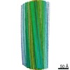 14187MC  7qvcC  7qvfC  7qwlC 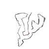 7qwmC M: map data used to model this data C: citing same article ( |
|---|---|
| Similar structure data |


-Links
 PDBj
PDBj
- Assembly
| Deposited unit | 
|
|---|---|
| 1 |
|
-Components
| #1: Protein | Mass: 31156.318 Da / Num. of mol.: 3 / Source method: isolated from a natural source / Source: (natural) Homo sapiens (human) / References: UniProt: Q9NUM4Has protein modification | Y | |
|---|
-Experimental details
-Experiment
| Experiment | Method: ELECTRON MICROSCOPY |
|---|---|
| EM experiment | Aggregation state: FILAMENT / 3D reconstruction method: helical reconstruction |
- Sample preparation
| Component | Name: TMEM106B / Type: TISSUE / Entity ID: all / Source: NATURAL |
|---|---|
| Source (natural) | Organism: Homo sapiens (human) |
| Buffer solution | pH: 7.4 |
| Specimen | Embedding applied: NO / Shadowing applied: NO / Staining applied: NO / Vitrification applied: YES |
| Vitrification | Cryogen name: ETHANE |
- Electron microscopy imaging
| Experimental equipment |  Model: Titan Krios / Image courtesy: FEI Company |
|---|---|
| Microscopy | Model: FEI TITAN KRIOS |
| Electron gun | Electron source:  FIELD EMISSION GUN / Accelerating voltage: 300 kV / Illumination mode: FLOOD BEAM FIELD EMISSION GUN / Accelerating voltage: 300 kV / Illumination mode: FLOOD BEAM |
| Electron lens | Mode: BRIGHT FIELD / Nominal defocus max: 3000 nm / Nominal defocus min: 1200 nm |
| Image recording | Electron dose: 40 e/Å2 / Film or detector model: FEI FALCON IV (4k x 4k) |
- Processing
| EM software |
| |||||||||||||||
|---|---|---|---|---|---|---|---|---|---|---|---|---|---|---|---|---|
| CTF correction | Type: PHASE FLIPPING AND AMPLITUDE CORRECTION | |||||||||||||||
| Helical symmerty | Angular rotation/subunit: -0.63 ° / Axial rise/subunit: 4.78 Å / Axial symmetry: C1 | |||||||||||||||
| 3D reconstruction | Resolution: 3.38 Å / Resolution method: FSC 0.143 CUT-OFF / Num. of particles: 7347 / Symmetry type: HELICAL |
