 Movie
Movie Controller


+ Open data

- Basic information
| Entry | Database: PDB / ID: 7qt9 | ||||||
|---|---|---|---|---|---|---|---|
| Title | Room temperature In-situ SARS-CoV-2 MPRO with bound Z4439011584 | ||||||
 Components Components | Non-structural protein 6 | ||||||
 Keywords Keywords | VIRAL PROTEIN / Protease / Main protease / SARS-CoV-2 | ||||||
| Function / homology |  Function and homology information Function and homology informationmain proteinase (3clpro) structure, domain 3 / main proteinase (3clpro) structure, domain 3 / Trypsin-like serine proteases / Thrombin, subunit H / Beta Barrel / Orthogonal Bundle / Mainly Beta / Mainly Alpha Similarity search - Domain/homology | ||||||
| Biological species |  Severe acute respiratory syndrome coronavirus Severe acute respiratory syndrome coronavirus | ||||||
| Method |  X-RAY DIFFRACTION / SYNCHROTRON / MOLECULAR REPLACEMENT / Resolution: 2.43 Å X-RAY DIFFRACTION / SYNCHROTRON / MOLECULAR REPLACEMENT / Resolution: 2.43 Å | ||||||
 Authors Authors | Horrell, S. / Gildae, R.J. / Axford, D. / Owen, C.D. / Lukacik, P. / Strain-Damerell, C. / Owen, R.L. / Walsh, M.A. | ||||||
| Funding support |  United Kingdom, 1items United Kingdom, 1items
| ||||||
 Citation Citation | Journal: Acta Crystallogr D Struct Biol / Year: 2022 Title: xia2.multiplex: a multi-crystal data-analysis pipeline. Authors: Gildea, R.J. / Beilsten-Edmands, J. / Axford, D. / Horrell, S. / Aller, P. / Sandy, J. / Sanchez-Weatherby, J. / Owen, C.D. / Lukacik, P. / Strain-Damerell, C. / Owen, R.L. / Walsh, M.A. / Winter, G. | ||||||
| History |
|
- Structure visualization
| Structure viewer | Molecule: MolmilJmol/JSmol |
|---|
- Downloads & links
-Download
| PDBx/mmCIF format | 7qt9.cif.gz | 225.6 KB | Display | PDBx/mmCIF format |
|---|---|---|---|---|
| PDB format | pdb7qt9.ent.gz | 176.6 KB | Display | PDB format |
| PDBx/mmJSON format | 7qt9.json.gz | Tree view | PDBx/mmJSON format | |
| Others |  Other downloads Other downloads |
-Validation report
| Arichive directory | https://data.pdbj.org/pub/pdb/validation_reports/qt/7qt9ftp://data.pdbj.org/pub/pdb/validation_reports/qt/7qt9 | HTTPS FTP |
|---|
-Related structure data
| Related structure data | 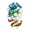 7qt5C 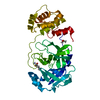 7qt6C 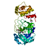 7qt7C  7qt8C 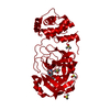 7aehS S: Starting model for refinement C: citing same article ( |
|---|---|
| Similar structure data | |
| Experimental dataset #1 | Data reference: 10.5281/zenodo.5836055 |
-Links
 PDBj
PDBj- Assembly
| Deposited unit | 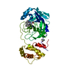
| ||||||||
|---|---|---|---|---|---|---|---|---|---|
| 1 |
| ||||||||
| Unit cell |
|
-Components
| #1: Protein | Mass: 33697.418 Da / Num. of mol.: 1 Source method: isolated from a genetically manipulated source Source: (gene. exp.) Severe acute respiratory syndrome coronavirusGene: ORF1ab / Production host:  |
|---|---|
| #2: Chemical | ChemComp-UJ1 / 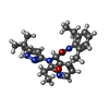  Mass: 447.573 Da / Num. of mol.: 1 / Source method: obtained synthetically / Formula: C26H33N5O2 / Feature type: SUBJECT OF INVESTIGATION Mass: 447.573 Da / Num. of mol.: 1 / Source method: obtained synthetically / Formula: C26H33N5O2 / Feature type: SUBJECT OF INVESTIGATION |
| #3: Chemical | ChemComp-DMS /   Mass: 78.133 Da / Num. of mol.: 1 / Source method: obtained synthetically / Formula: C2H6OS / Comment: DMSO, precipitant*YM Mass: 78.133 Da / Num. of mol.: 1 / Source method: obtained synthetically / Formula: C2H6OS / Comment: DMSO, precipitant*YM |
| #4: Water | ChemComp-HOH /  Mass: 18.015 Da / Num. of mol.: 23 / Source method: isolated from a natural source / Formula: H2O Mass: 18.015 Da / Num. of mol.: 23 / Source method: isolated from a natural source / Formula: H2O |
| Has ligand of interest | Y |
| Has protein modification | Y |
-Experimental details
-Experiment
| Experiment | Method: X-RAY DIFFRACTION / Number of used crystals: 1 |
|---|
- Sample preparation
| Crystal | Density Matthews: 2.07 Å3/Da / Density % sol: 40.72 % |
|---|---|
| Crystal grow | Temperature: 298 K / Method: vapor diffusion / pH: 6.5 / Details: 11% (v/v) PEG 4K, 5% (v/v) DMSO, 0.1 M MES pH 6.5 |
-Data collection
| Diffraction | Mean temperature: 298 K / Serial crystal experiment: N |
|---|---|
| Diffraction source | Source: SYNCHROTRON / Site: Diamond / Beamline: I24 / Wavelength: 1 Å |
| Detector | Type: DECTRIS PILATUS 6M-F / Detector: PIXEL / Date: May 6, 2020 |
| Radiation | Protocol: SINGLE WAVELENGTH / Monochromatic (M) / Laue (L): M / Scattering type: x-ray |
| Radiation wavelength | Wavelength: 1 Å / Relative weight: 1 |
| Reflection | Resolution: 2.43→56.846 Å / Num. obs: 10345 / % possible obs: 97.9 % / Redundancy: 5.9 % / CC1/2: 0.987 / Rmerge(I) obs: 0.166 / Rpim(I) all: 0.073 / Rsym value: 0.183 / Net I/σ(I): 10.8 |
| Reflection shell | Resolution: 2.43→2.52 Å / Redundancy: 5.9 % / Rmerge(I) obs: 1.241 / Num. unique obs: 1038 / CC1/2: 0.305 / Rpim(I) all: 0.578 / Rrim(I) all: 1.38 / % possible all: 97.7 |
- Processing
| Software |
| ||||||||||||||||||||||||||||||||||||||||||||||||||||||||||||||||||||||||||||||||||||||||||||||||||||||||||||||||||||||||||||||||||||||||||||||||||||||||||||||||
|---|---|---|---|---|---|---|---|---|---|---|---|---|---|---|---|---|---|---|---|---|---|---|---|---|---|---|---|---|---|---|---|---|---|---|---|---|---|---|---|---|---|---|---|---|---|---|---|---|---|---|---|---|---|---|---|---|---|---|---|---|---|---|---|---|---|---|---|---|---|---|---|---|---|---|---|---|---|---|---|---|---|---|---|---|---|---|---|---|---|---|---|---|---|---|---|---|---|---|---|---|---|---|---|---|---|---|---|---|---|---|---|---|---|---|---|---|---|---|---|---|---|---|---|---|---|---|---|---|---|---|---|---|---|---|---|---|---|---|---|---|---|---|---|---|---|---|---|---|---|---|---|---|---|---|---|---|---|---|---|---|---|
| Refinement | Method to determine structure: MOLECULAR REPLACEMENT Starting model: 7AEH Resolution: 2.43→56.846 Å / Cor.coef. Fo:Fc: 0.975 / Cor.coef. Fo:Fc free: 0.953 / SU B: 21.705 / SU ML: 0.207 / Cross valid method: FREE R-VALUE / ESU R: 1.571 / ESU R Free: 0.248 Details: Hydrogens have been added in their riding positions
| ||||||||||||||||||||||||||||||||||||||||||||||||||||||||||||||||||||||||||||||||||||||||||||||||||||||||||||||||||||||||||||||||||||||||||||||||||||||||||||||||
| Solvent computation | Ion probe radii: 0.8 Å / Shrinkage radii: 0.8 Å / VDW probe radii: 1.2 Å / Solvent model: MASK BULK SOLVENT | ||||||||||||||||||||||||||||||||||||||||||||||||||||||||||||||||||||||||||||||||||||||||||||||||||||||||||||||||||||||||||||||||||||||||||||||||||||||||||||||||
| Displacement parameters | Biso mean: 45.722 Å2
| ||||||||||||||||||||||||||||||||||||||||||||||||||||||||||||||||||||||||||||||||||||||||||||||||||||||||||||||||||||||||||||||||||||||||||||||||||||||||||||||||
| Refinement step | Cycle: LAST / Resolution: 2.43→56.846 Å
| ||||||||||||||||||||||||||||||||||||||||||||||||||||||||||||||||||||||||||||||||||||||||||||||||||||||||||||||||||||||||||||||||||||||||||||||||||||||||||||||||
| Refine LS restraints |
| ||||||||||||||||||||||||||||||||||||||||||||||||||||||||||||||||||||||||||||||||||||||||||||||||||||||||||||||||||||||||||||||||||||||||||||||||||||||||||||||||
| LS refinement shell |
| ||||||||||||||||||||||||||||||||||||||||||||||||||||||||||||||||||||||||||||||||||||||||||||||||||||||||||||||||||||||||||||||||||||||||||||||||||||||||||||||||
| Refinement TLS params. | Method: refined / Origin x: -2.1659 Å / Origin y: 0.7661 Å / Origin z: 13.0578 Å
| ||||||||||||||||||||||||||||||||||||||||||||||||||||||||||||||||||||||||||||||||||||||||||||||||||||||||||||||||||||||||||||||||||||||||||||||||||||||||||||||||
| Refinement TLS group | Selection: ALL |
