 Movie
Movie Controller

[English] 日本語
 Yorodumi
- PDB-7qql: The PDZ domain of SNTG2 complexed with the phosphorylated PDZ-bin... -
+ Open data

- Basic information
| Entry | Database: PDB / ID: 7qql | ||||||
|---|---|---|---|---|---|---|---|
| Title | The PDZ domain of SNTG2 complexed with the phosphorylated PDZ-binding motif of RSK1 | ||||||
 Components Components |
| ||||||
 Keywords Keywords | PEPTIDE BINDING PROTEIN / PDZ / linear motif / crystallization chaperone | ||||||
| Function / homology |  Function and homology information Function and homology informationsyntrophin complex / regulation of translation in response to stress / CREB1 phosphorylation through NMDA receptor-mediated activation of RAS signaling / AnxA2-p11 complex / membrane raft assembly / positive regulation of receptor-mediated endocytosis involved in cholesterol transport / positive regulation of vacuole organization / dystrophin-associated glycoprotein complex / ribosomal protein S6 kinase activity / phospholipase A2 inhibitor activity ...syntrophin complex / regulation of translation in response to stress / CREB1 phosphorylation through NMDA receptor-mediated activation of RAS signaling / AnxA2-p11 complex / membrane raft assembly / positive regulation of receptor-mediated endocytosis involved in cholesterol transport / positive regulation of vacuole organization / dystrophin-associated glycoprotein complex / ribosomal protein S6 kinase activity / phospholipase A2 inhibitor activity / positive regulation of low-density lipoprotein particle clearance / positive regulation of vesicle fusion / hepatocyte proliferation / CREB phosphorylation / myelin sheath adaxonal region / positive regulation of hepatic stellate cell activation / negative regulation of low-density lipoprotein particle receptor catabolic process / neuroligin family protein binding / positive regulation of plasma membrane repair / positive regulation of plasminogen activation / PCSK9-AnxA2 complex / cadherin binding involved in cell-cell adhesion / cornified envelope / Schmidt-Lanterman incisure / vesicle budding from membrane / Gastrin-CREB signalling pathway via PKC and MAPK / TORC1 signaling / calcium-dependent phospholipid binding / negative regulation of receptor internalization / plasma membrane protein complex / osteoclast development / RSK activation / Dissolution of Fibrin Clot / S100 protein binding / collagen fibril organization / negative regulation of TOR signaling / vesicle membrane / epithelial cell apoptotic process / phosphatidylserine binding / ERK/MAPK targets / Recycling pathway of L1 / positive regulation of receptor recycling / basement membrane / positive regulation of exocytosis / Smooth Muscle Contraction / regulation of neurogenesis / fibrinolysis / cytoskeletal protein binding / phosphatidylinositol-4,5-bisphosphate binding / lipid droplet / Transcriptional and post-translational regulation of MITF-M expression and activity / protein serine/threonine/tyrosine kinase activity / Gene and protein expression by JAK-STAT signaling after Interleukin-12 stimulation / lung development / Turbulent (oscillatory, disturbed) flow shear stress activates signaling by PIEZO1 and integrins in endothelial cells / cell-matrix adhesion / central nervous system development / response to activity / PDZ domain binding / adherens junction / positive regulation of cell differentiation / serine-type endopeptidase inhibitor activity / mRNA transcription by RNA polymerase II / sarcolemma / RNA polymerase II transcription regulator complex / nuclear matrix / calcium-dependent protein binding / : / azurophil granule lumen / late endosome membrane / melanosome / actin binding / positive regulation of cell growth / protease binding / Senescence-Associated Secretory Phenotype (SASP) / angiogenesis / midbody / basolateral plasma membrane / vesicle / chemical synaptic transmission / early endosome / cytoskeleton / protein phosphorylation / non-specific serine/threonine protein kinase / endosome / lysosomal membrane / protein serine kinase activity / protein serine/threonine kinase activity / calcium ion binding / Neutrophil degranulation / synapse / negative regulation of apoptotic process / positive regulation of DNA-templated transcription / structural molecule activity / cell surface / magnesium ion binding / signal transduction / positive regulation of transcription by RNA polymerase II / extracellular space / RNA binding Similarity search - Function | ||||||
| Biological species |  Homo sapiens (human) Homo sapiens (human) | ||||||
| Method |  X-RAY DIFFRACTION / SYNCHROTRON / MOLECULAR REPLACEMENT / Resolution: 2.44 Å X-RAY DIFFRACTION / SYNCHROTRON / MOLECULAR REPLACEMENT / Resolution: 2.44 Å | ||||||
 Authors Authors | Cousido-Siah, A. / Trave, G. / Gogl, G. | ||||||
| Funding support | 1items
| ||||||
 Citation Citation | Journal: Acta Crystallogr D Struct Biol / Year: 2022 Title: A scalable strategy to solve structures of PDZ domains and their complexes. Authors: Cousido-Siah, A. / Carneiro, L. / Kostmann, C. / Ecsedi, P. / Nyitray, L. / Trave, G. / Gogl, G. | ||||||
| History |
|
- Structure visualization
| Structure viewer | Molecule: MolmilJmol/JSmol |
|---|
- Downloads & links
-Download
| PDBx/mmCIF format | 7qql.cif.gz | 495.4 KB | Display | PDBx/mmCIF format |
|---|---|---|---|---|
| PDB format | pdb7qql.ent.gz | 408.6 KB | Display | PDB format |
| PDBx/mmJSON format | 7qql.json.gz | Tree view | PDBx/mmJSON format | |
| Others |  Other downloads Other downloads |
-Validation report
| Arichive directory | https://data.pdbj.org/pub/pdb/validation_reports/qq/7qqlftp://data.pdbj.org/pub/pdb/validation_reports/qq/7qql | HTTPS FTP |
|---|
-Related structure data
| Related structure data |  7pc3C  7pc4C  7pc5C  7pc7C  7pc8C 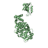 7pc9C 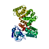 7pcbC 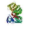 7qqmC 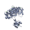 7qqnC  5n7dS S: Starting model for refinement C: citing same article ( |
|---|---|
| Similar structure data |
-Links
 PDBj
PDBj


- Assembly
| Deposited unit | 
| ||||||||
|---|---|---|---|---|---|---|---|---|---|
| 1 | 
| ||||||||
| 2 | 
| ||||||||
| 3 | 
| ||||||||
| Unit cell |
|
-Components
| #1: Protein | Mass: 46138.352 Da / Num. of mol.: 3 Source method: isolated from a genetically manipulated source Source: (gene. exp.) Homo sapiens (human) / Gene: SNTG2, ANXA2, ANX2, ANX2L4, CAL1H, LPC2D / Production host:  #2: Protein/peptide | Mass: 1410.602 Da / Num. of mol.: 3 / Source method: obtained synthetically Details: biotin-ttds (Trioxatridecan-succinamic acid) tag at the N-terminus Source: (synth.) Homo sapiens (human)References: UniProt: Q15418, non-specific serine/threonine protein kinase #3: Chemical | ChemComp-CA /   Mass: 40.078 Da / Num. of mol.: 14 / Source method: isolated from a natural source / Formula: Ca Mass: 40.078 Da / Num. of mol.: 14 / Source method: isolated from a natural source / Formula: Ca#4: Chemical |   Mass: 92.094 Da / Num. of mol.: 3 / Source method: obtained synthetically / Formula: C3H8O3 Mass: 92.094 Da / Num. of mol.: 3 / Source method: obtained synthetically / Formula: C3H8O3#5: Water | ChemComp-HOH / |  Mass: 18.015 Da / Num. of mol.: 223 / Source method: isolated from a natural source / Formula: H2O Mass: 18.015 Da / Num. of mol.: 223 / Source method: isolated from a natural source / Formula: H2OHas ligand of interest | Y | Has protein modification | Y | |
|---|
-Experimental details
-Experiment
| Experiment | Method: X-RAY DIFFRACTION / Number of used crystals: 1 |
|---|
- Sample preparation
| Crystal | Density Matthews: 2.81 Å3/Da / Density % sol: 56.17 % |
|---|---|
| Crystal grow | Temperature: 298 K / Method: vapor diffusion, sitting drop / Details: 0.1 M Succinic acid pH 7.0 15% w/v PEG 3350 |
-Data collection
| Diffraction | Mean temperature: 100 K / Serial crystal experiment: N |
|---|---|
| Diffraction source | Source: SYNCHROTRON / Site: SLS  / Beamline: X06DA / Wavelength: 1 Å / Beamline: X06DA / Wavelength: 1 Å |
| Detector | Type: DECTRIS PILATUS 2M / Detector: PIXEL / Date: Oct 25, 2021 |
| Radiation | Protocol: SINGLE WAVELENGTH / Monochromatic (M) / Laue (L): M / Scattering type: x-ray |
| Radiation wavelength | Wavelength: 1 Å / Relative weight: 1 |
| Reflection | Resolution: 2.44→48.416 Å / Num. obs: 59163 / % possible obs: 99.9 % / Redundancy: 6.79 % / CC1/2: 0.998 / Rrim(I) all: 0.147 / Net I/σ(I): 11.3 |
| Reflection shell | Resolution: 2.44→2.5 Å / Mean I/σ(I) obs: 1.18 / Num. unique obs: 4350 / CC1/2: 0.454 / Rrim(I) all: 1.655 |
- Processing
| Software |
| ||||||||||||||||||||||||||||||||||||||||||||||||||||||||||||||||||||||||||||||||||||||||||||||||||||||||||||||||||||||||||||||||||||||||||||||||||||||||||||||||||||||||||||||||||||||||||||||||||||||||||||||||||||||||||||||||||||||||||||||||||||||||||||||||||||||||||||||||||||||||||||||||||||||||||||||||||||||||||||||||||||||||||||||||||||||||||||||
|---|---|---|---|---|---|---|---|---|---|---|---|---|---|---|---|---|---|---|---|---|---|---|---|---|---|---|---|---|---|---|---|---|---|---|---|---|---|---|---|---|---|---|---|---|---|---|---|---|---|---|---|---|---|---|---|---|---|---|---|---|---|---|---|---|---|---|---|---|---|---|---|---|---|---|---|---|---|---|---|---|---|---|---|---|---|---|---|---|---|---|---|---|---|---|---|---|---|---|---|---|---|---|---|---|---|---|---|---|---|---|---|---|---|---|---|---|---|---|---|---|---|---|---|---|---|---|---|---|---|---|---|---|---|---|---|---|---|---|---|---|---|---|---|---|---|---|---|---|---|---|---|---|---|---|---|---|---|---|---|---|---|---|---|---|---|---|---|---|---|---|---|---|---|---|---|---|---|---|---|---|---|---|---|---|---|---|---|---|---|---|---|---|---|---|---|---|---|---|---|---|---|---|---|---|---|---|---|---|---|---|---|---|---|---|---|---|---|---|---|---|---|---|---|---|---|---|---|---|---|---|---|---|---|---|---|---|---|---|---|---|---|---|---|---|---|---|---|---|---|---|---|---|---|---|---|---|---|---|---|---|---|---|---|---|---|---|---|---|---|---|---|---|---|---|---|---|---|---|---|---|---|---|---|---|---|---|---|---|---|---|---|---|---|---|---|---|---|---|---|---|---|---|---|---|---|---|---|---|---|---|---|---|---|---|---|---|---|---|---|---|---|---|---|---|---|---|---|---|---|---|---|---|---|---|---|---|---|---|---|---|---|---|---|---|---|---|---|---|---|---|---|
| Refinement | Method to determine structure: MOLECULAR REPLACEMENT Starting model: 5N7D Resolution: 2.44→48.416 Å / SU ML: 0.3 / Cross valid method: THROUGHOUT / σ(F): 1.36 / Phase error: 24.37 / Stereochemistry target values: ML
| ||||||||||||||||||||||||||||||||||||||||||||||||||||||||||||||||||||||||||||||||||||||||||||||||||||||||||||||||||||||||||||||||||||||||||||||||||||||||||||||||||||||||||||||||||||||||||||||||||||||||||||||||||||||||||||||||||||||||||||||||||||||||||||||||||||||||||||||||||||||||||||||||||||||||||||||||||||||||||||||||||||||||||||||||||||||||||||||
| Solvent computation | Shrinkage radii: 0.9 Å / VDW probe radii: 1.11 Å / Solvent model: FLAT BULK SOLVENT MODEL | ||||||||||||||||||||||||||||||||||||||||||||||||||||||||||||||||||||||||||||||||||||||||||||||||||||||||||||||||||||||||||||||||||||||||||||||||||||||||||||||||||||||||||||||||||||||||||||||||||||||||||||||||||||||||||||||||||||||||||||||||||||||||||||||||||||||||||||||||||||||||||||||||||||||||||||||||||||||||||||||||||||||||||||||||||||||||||||||
| Displacement parameters | Biso max: 215.35 Å2 / Biso mean: 66.7492 Å2 / Biso min: 26.31 Å2 | ||||||||||||||||||||||||||||||||||||||||||||||||||||||||||||||||||||||||||||||||||||||||||||||||||||||||||||||||||||||||||||||||||||||||||||||||||||||||||||||||||||||||||||||||||||||||||||||||||||||||||||||||||||||||||||||||||||||||||||||||||||||||||||||||||||||||||||||||||||||||||||||||||||||||||||||||||||||||||||||||||||||||||||||||||||||||||||||
| Refinement step | Cycle: final / Resolution: 2.44→48.416 Å
| ||||||||||||||||||||||||||||||||||||||||||||||||||||||||||||||||||||||||||||||||||||||||||||||||||||||||||||||||||||||||||||||||||||||||||||||||||||||||||||||||||||||||||||||||||||||||||||||||||||||||||||||||||||||||||||||||||||||||||||||||||||||||||||||||||||||||||||||||||||||||||||||||||||||||||||||||||||||||||||||||||||||||||||||||||||||||||||||
| LS refinement shell | Refine-ID: X-RAY DIFFRACTION / Rfactor Rfree error: 0 / % reflection obs: 100 %
| ||||||||||||||||||||||||||||||||||||||||||||||||||||||||||||||||||||||||||||||||||||||||||||||||||||||||||||||||||||||||||||||||||||||||||||||||||||||||||||||||||||||||||||||||||||||||||||||||||||||||||||||||||||||||||||||||||||||||||||||||||||||||||||||||||||||||||||||||||||||||||||||||||||||||||||||||||||||||||||||||||||||||||||||||||||||||||||||
| Refinement TLS params. | Method: refined / Refine-ID: X-RAY DIFFRACTION
| ||||||||||||||||||||||||||||||||||||||||||||||||||||||||||||||||||||||||||||||||||||||||||||||||||||||||||||||||||||||||||||||||||||||||||||||||||||||||||||||||||||||||||||||||||||||||||||||||||||||||||||||||||||||||||||||||||||||||||||||||||||||||||||||||||||||||||||||||||||||||||||||||||||||||||||||||||||||||||||||||||||||||||||||||||||||||||||||
| Refinement TLS group |
|