 Movie
Movie Controller

[English] 日本語
 Yorodumi
- PDB-7qpu: Botulinum neurotoxin A5 cell binding domain in complex with GM1b ... -
+ Open data

- Basic information
| Entry | Database: PDB / ID: 7qpu | ||||||
|---|---|---|---|---|---|---|---|
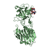
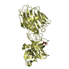
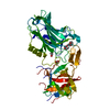
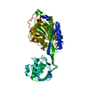
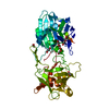
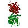
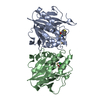
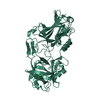
| Title | Botulinum neurotoxin A5 cell binding domain in complex with GM1b oligosaccharide | ||||||
 Components Components | Botulinum neurotoxin sub-type A5 | ||||||
 Keywords Keywords | TOXIN / Cell binding domain / receptor / oligosaccharide / neurotoxin | ||||||
| Function / homology |  Function and homology information Function and homology informationbontoxilysin / protein transmembrane transporter activity / metalloendopeptidase activity / toxin activity / proteolysis / extracellular region / zinc ion binding Similarity search - Function | ||||||
| Biological species |   Clostridium botulinum (bacteria) Clostridium botulinum (bacteria) | ||||||
| Method |  X-RAY DIFFRACTION / SYNCHROTRON / MOLECULAR REPLACEMENT / Resolution: 2.4 Å X-RAY DIFFRACTION / SYNCHROTRON / MOLECULAR REPLACEMENT / Resolution: 2.4 Å | ||||||
 Authors Authors | Gregory, K.S. / Acharya, K.R. / Liu, S.M. | ||||||
| Funding support | 1items
| ||||||
 Citation Citation | Journal: Toxins / Year: 2022 Title: Crystal Structures of Botulinum Neurotoxin Subtypes A4 and A5 Cell Binding Domains in Complex with Receptor Ganglioside. Authors: Gregory, K.S. / Mojanaga, O.O. / Liu, S.M. / Acharya, K.R. | ||||||
| History |
|
- Structure visualization
| Structure viewer | Molecule: MolmilJmol/JSmol |
|---|
- Downloads & links
-Download
| PDBx/mmCIF format | 7qpu.cif.gz | 208.3 KB | Display | PDBx/mmCIF format |
|---|---|---|---|---|
| PDB format | pdb7qpu.ent.gz | 131.5 KB | Display | PDB format |
| PDBx/mmJSON format | 7qpu.json.gz | Tree view | PDBx/mmJSON format | |
| Others |  Other downloads Other downloads |
-Validation report
| Arichive directory | https://data.pdbj.org/pub/pdb/validation_reports/qp/7qpuftp://data.pdbj.org/pub/pdb/validation_reports/qp/7qpu | HTTPS FTP |
|---|
-Related structure data
| Related structure data | 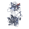 7qptC 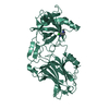 6twpS S: Starting model for refinement C: citing same article ( |
|---|---|
| Similar structure data |

-Links
 PDBj
PDBj- Assembly
| Deposited unit | 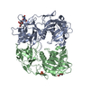
| ||||||||||||
|---|---|---|---|---|---|---|---|---|---|---|---|---|---|
| 1 | 
| ||||||||||||
| 2 | 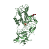
| ||||||||||||
| Unit cell |
|
-Components
| #1: Protein | Mass: 50719.418 Da / Num. of mol.: 2 Source method: isolated from a genetically manipulated source Source: (gene. exp.) Clostridium botulinum (bacteria) / Gene: cntA, cntA/A5 / Production host: #2: Polysaccharide | N-acetyl-alpha-neuraminic acid-(2-3)-beta-D-galactopyranose | Source method: isolated from a genetically manipulated source #3: Chemical | ChemComp-PEG / |   Mass: 106.120 Da / Num. of mol.: 1 / Source method: obtained synthetically / Formula: C4H10O3 Mass: 106.120 Da / Num. of mol.: 1 / Source method: obtained synthetically / Formula: C4H10O3#4: Chemical | ChemComp-SO4 / |   Mass: 96.063 Da / Num. of mol.: 1 / Source method: obtained synthetically / Formula: SO4 Mass: 96.063 Da / Num. of mol.: 1 / Source method: obtained synthetically / Formula: SO4#5: Water | ChemComp-HOH / |  Mass: 18.015 Da / Num. of mol.: 165 / Source method: isolated from a natural source / Formula: H2O Mass: 18.015 Da / Num. of mol.: 165 / Source method: isolated from a natural source / Formula: H2OHas ligand of interest | Y | |
|---|
-Experimental details
-Experiment
| Experiment | Method: X-RAY DIFFRACTION / Number of used crystals: 1 |
|---|
- Sample preparation
| Crystal | Density Matthews: 2.17 Å3/Da / Density % sol: 43.45 % |
|---|---|
| Crystal grow | Temperature: 289.15 K / Method: vapor diffusion, sitting drop / pH: 7.8 Details: 150 mM Li2SO4, 50 mM MgCl2 6H2O , 0.1 M HEPES pH 7.8, 4.7% w/v PEG 8K, 4.7% PEG 10K and 4.7% PEG 8K |
-Data collection
| Diffraction | Mean temperature: 100 K / Serial crystal experiment: N |
|---|---|
| Diffraction source | Source: SYNCHROTRON / Site: Diamond  / Beamline: I04 / Wavelength: 0.9795 Å / Beamline: I04 / Wavelength: 0.9795 Å |
| Detector | Type: DECTRIS EIGER2 X 16M / Detector: PIXEL / Date: Mar 2, 2020 |
| Radiation | Protocol: SINGLE WAVELENGTH / Monochromatic (M) / Laue (L): M / Scattering type: x-ray |
| Radiation wavelength | Wavelength: 0.9795 Å / Relative weight: 1 |
| Reflection | Resolution: 2.4→129.4 Å / Num. obs: 33451 / % possible obs: 100 % / Redundancy: 6.8 % / Biso Wilson estimate: 36.33 Å2 / CC1/2: 0.982 / Rpim(I) all: 0.126 / Net I/σ(I): 4.3 |
| Reflection shell | Resolution: 2.4→2.49 Å / Num. unique obs: 22543 / CC1/2: 0.575 / Rpim(I) all: 0.746 |
- Processing
| Software |
| ||||||||||||||||||||||||||||||||||||||||||||||||||||||||||||||||||||||||||||||||||||
|---|---|---|---|---|---|---|---|---|---|---|---|---|---|---|---|---|---|---|---|---|---|---|---|---|---|---|---|---|---|---|---|---|---|---|---|---|---|---|---|---|---|---|---|---|---|---|---|---|---|---|---|---|---|---|---|---|---|---|---|---|---|---|---|---|---|---|---|---|---|---|---|---|---|---|---|---|---|---|---|---|---|---|---|---|---|
| Refinement | Method to determine structure: MOLECULAR REPLACEMENT Starting model: 6TWP Resolution: 2.4→76.07 Å / SU ML: 0.3514 / Cross valid method: FREE R-VALUE / σ(F): 1.34 / Phase error: 28.2652 Stereochemistry target values: GeoStd + Monomer Library + CDL v1.2
| ||||||||||||||||||||||||||||||||||||||||||||||||||||||||||||||||||||||||||||||||||||
| Solvent computation | Shrinkage radii: 0.9 Å / VDW probe radii: 1.11 Å / Solvent model: FLAT BULK SOLVENT MODEL | ||||||||||||||||||||||||||||||||||||||||||||||||||||||||||||||||||||||||||||||||||||
| Displacement parameters | Biso mean: 38.72 Å2 | ||||||||||||||||||||||||||||||||||||||||||||||||||||||||||||||||||||||||||||||||||||
| Refinement step | Cycle: LAST / Resolution: 2.4→76.07 Å
| ||||||||||||||||||||||||||||||||||||||||||||||||||||||||||||||||||||||||||||||||||||
| Refine LS restraints |
| ||||||||||||||||||||||||||||||||||||||||||||||||||||||||||||||||||||||||||||||||||||
| LS refinement shell |
|
