 Movie
Movie Controller


+ Open data

- Basic information
| Entry | Database: PDB / ID: 7qpr | ||||||
|---|---|---|---|---|---|---|---|
| Title | Structure of full length SpoT | ||||||
 Components Components | ACT domain protein | ||||||
 Keywords Keywords | HYDROLASE / (p)ppGpp hydrolase bound to ppGpp | ||||||
| Function / homology |  Function and homology information Function and homology informationguanosine-3',5'-bis(diphosphate) 3'-diphosphatase / guanosine-3',5'-bis(diphosphate) 3'-diphosphatase activity / GTP diphosphokinase activity / guanosine tetraphosphate biosynthetic process / response to starvation / kinase activity / nucleotide binding / metal ion binding / plasma membrane Similarity search - Function | ||||||
| Biological species |  Acinetobacter baumannii (bacteria) Acinetobacter baumannii (bacteria) | ||||||
| Method |  X-RAY DIFFRACTION / SYNCHROTRON / MOLECULAR REPLACEMENT / Resolution: 2.513 Å X-RAY DIFFRACTION / SYNCHROTRON / MOLECULAR REPLACEMENT / Resolution: 2.513 Å | ||||||
 Authors Authors | Garcia-Pino, A. / Tamman, H. | ||||||
| Funding support | 1items
| ||||||
 Citation Citation | Journal: To Be Published Title: Structure of full length SpoT Authors: Garcia-Pino, A. / Tamman, H. | ||||||
| History |
|
- Structure visualization
| Structure viewer | Molecule: MolmilJmol/JSmol |
|---|
- Downloads & links
-Download
| PDBx/mmCIF format | 7qpr.cif.gz | 1.1 MB | Display | PDBx/mmCIF format |
|---|---|---|---|---|
| PDB format | pdb7qpr.ent.gz | 905.8 KB | Display | PDB format |
| PDBx/mmJSON format | 7qpr.json.gz | Tree view | PDBx/mmJSON format | |
| Others |  Other downloads Other downloads |
-Validation report
| Arichive directory | https://data.pdbj.org/pub/pdb/validation_reports/qp/7qprftp://data.pdbj.org/pub/pdb/validation_reports/qp/7qpr | HTTPS FTP |
|---|
-Related structure data
| Related structure data | 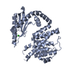 6s2vS S: Starting model for refinement |
|---|---|
| Similar structure data |
-Links
 PDBj
PDBj


- Assembly
| Deposited unit | 
| ||||||||
|---|---|---|---|---|---|---|---|---|---|
| 1 | 
| ||||||||
| 2 | 
| ||||||||
| 3 | 
| ||||||||
| 4 | 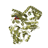
| ||||||||
| Unit cell |
|
-Components
-Protein , 1 types, 4 molecules ABCD
| #1: Protein | Mass: 79791.141 Da / Num. of mol.: 4 Source method: isolated from a genetically manipulated source Source: (gene. exp.) Acinetobacter baumannii (bacteria)Gene: spoT_1, relA_1, relA_3, spoT, A7M90_14410, AB945B12_01945, Aba9201_15290, ABCAM1_3401, ABKPCSM17A_01292, ABR2091_3374, ABUW_0309, APC21_16100, APD31_02670, AUO97_04875, AYR68_16515, B7L36_ ...Gene: spoT_1, relA_1, relA_3, spoT, A7M90_14410, AB945B12_01945, Aba9201_15290, ABCAM1_3401, ABKPCSM17A_01292, ABR2091_3374, ABUW_0309, APC21_16100, APD31_02670, AUO97_04875, AYR68_16515, B7L36_04505, B7L45_01975, B9X95_19475, BAA1790NC_0315, BS065_01600, C2U32_12605, C5H40_03430, C6N18_18350, CBE85_06420, CBL15_01550, CSB70_3370, CTZ19_01585, DLI71_12465, DLI72_03665, DOL94_17785, E1A86_17370, E2535_18555, E2539_03475, E2540_19450, EA686_01605, EA706_01570, EA720_012255, EA722_01135, EGM95_01775, EKS29_00960, EP550_01655, EP560_16360, EWO96_11255, F2P40_01005, F4T85_13595, F4T91_15305, FDN00_18830, FE003_01590, FJU36_10385, FJU42_02940, FJU59_00430, FJU76_08505, FR761_17725, GNY86_13750, GSE42_18470, H0529_17955, H1058_16860, HBK86_16410, HIN86_01600, IMO23_16495, NCTC13305_02770, NCTC13421_00317, SAMEA104305281_00293, SAMEA104305340_01315, SAMEA104305385_01516, SI89_16095 Production host: References: UniProt: V5V8V7, GTP diphosphokinase |
|---|
-Non-polymers , 8 types, 895 molecules 

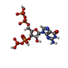


| #2: Chemical | ChemComp-MN /  Mass: 54.938 Da / Num. of mol.: 4 / Source method: obtained synthetically / Formula: Mn / Feature type: SUBJECT OF INVESTIGATION Mass: 54.938 Da / Num. of mol.: 4 / Source method: obtained synthetically / Formula: Mn / Feature type: SUBJECT OF INVESTIGATION#3: Chemical | ChemComp-ZN /  Mass: 65.409 Da / Num. of mol.: 4 / Source method: obtained synthetically / Formula: Zn / Feature type: SUBJECT OF INVESTIGATION Mass: 65.409 Da / Num. of mol.: 4 / Source method: obtained synthetically / Formula: Zn / Feature type: SUBJECT OF INVESTIGATION#4: Chemical | ChemComp-G4P /  Type: RNA linking / Mass: 603.160 Da / Num. of mol.: 4 / Source method: obtained synthetically / Formula: C10H17N5O17P4 / Feature type: SUBJECT OF INVESTIGATION Type: RNA linking / Mass: 603.160 Da / Num. of mol.: 4 / Source method: obtained synthetically / Formula: C10H17N5O17P4 / Feature type: SUBJECT OF INVESTIGATION#5: Chemical |  Mass: 189.100 Da / Num. of mol.: 2 / Source method: obtained synthetically / Formula: C6H5O7 Mass: 189.100 Da / Num. of mol.: 2 / Source method: obtained synthetically / Formula: C6H5O7#6: Chemical | ChemComp-GOL /  Mass: 92.094 Da / Num. of mol.: 4 / Source method: obtained synthetically / Formula: C3H8O3 Mass: 92.094 Da / Num. of mol.: 4 / Source method: obtained synthetically / Formula: C3H8O3#7: Chemical |  Mass: 35.453 Da / Num. of mol.: 3 / Source method: obtained synthetically / Formula: Cl Mass: 35.453 Da / Num. of mol.: 3 / Source method: obtained synthetically / Formula: Cl#8: Chemical |  Mass: 24.305 Da / Num. of mol.: 2 / Source method: obtained synthetically / Formula: Mg Mass: 24.305 Da / Num. of mol.: 2 / Source method: obtained synthetically / Formula: Mg#9: Water | ChemComp-HOH / | Mass: 18.015 Da / Num. of mol.: 872 / Source method: isolated from a natural source / Formula: H2O |
|---|
-Details
| Has ligand of interest | Y |
|---|
-Experimental details
-Experiment
| Experiment | Method: X-RAY DIFFRACTION / Number of used crystals: 1 |
|---|
- Sample preparation
| Crystal | Density Matthews: 2.91 Å3/Da / Density % sol: 57.77 % |
|---|---|
| Crystal grow | Temperature: 277 K / Method: vapor diffusion, sitting drop Details: 0.85M sodium citrate, 0.1M sodium chloride, 0.1M tris PH range: 8 |
-Data collection
| Diffraction | Mean temperature: 100 K / Serial crystal experiment: N |
|---|---|
| Diffraction source | Source: SYNCHROTRON / Site: SOLEIL  / Beamline: PROXIMA 1 / Wavelength: 0.9786 Å / Beamline: PROXIMA 1 / Wavelength: 0.9786 Å |
| Detector | Type: DECTRIS EIGER X 16M / Detector: PIXEL / Date: Nov 11, 2020 |
| Radiation | Protocol: SINGLE WAVELENGTH / Monochromatic (M) / Laue (L): M / Scattering type: x-ray |
| Radiation wavelength | Wavelength: 0.9786 Å / Relative weight: 1 |
| Reflection | Resolution: 2.513→48.88 Å / Num. obs: 83804 / % possible obs: 95.5 % / Redundancy: 13.8 % / Biso Wilson estimate: 74.68 Å2 / CC1/2: 0.995 / Rmerge(I) obs: 0.18 / Rpim(I) all: 0.051 / Net I/σ(I): 9.6 |
| Reflection shell | Resolution: 2.513→2.78 Å / Redundancy: 14 % / Rmerge(I) obs: 1.664 / Num. unique obs: 4191 / CC1/2: 0.712 / Rpim(I) all: 0.459 / % possible all: 72.6 |
- Processing
| Software |
| |||||||||||||||||||||||||||||||||||||||||||||||||||||||||||||||||||||||||||||||||||||||||||||||||||||||||||||||||||||||||||||
|---|---|---|---|---|---|---|---|---|---|---|---|---|---|---|---|---|---|---|---|---|---|---|---|---|---|---|---|---|---|---|---|---|---|---|---|---|---|---|---|---|---|---|---|---|---|---|---|---|---|---|---|---|---|---|---|---|---|---|---|---|---|---|---|---|---|---|---|---|---|---|---|---|---|---|---|---|---|---|---|---|---|---|---|---|---|---|---|---|---|---|---|---|---|---|---|---|---|---|---|---|---|---|---|---|---|---|---|---|---|---|---|---|---|---|---|---|---|---|---|---|---|---|---|---|---|---|
| Refinement | Method to determine structure: MOLECULAR REPLACEMENT Starting model: 6S2V Resolution: 2.513→48.88 Å / Cor.coef. Fo:Fc: 0.932 / Cor.coef. Fo:Fc free: 0.918 / Cross valid method: THROUGHOUT / σ(F): 0 / SU Rfree Blow DPI: 0.379
| |||||||||||||||||||||||||||||||||||||||||||||||||||||||||||||||||||||||||||||||||||||||||||||||||||||||||||||||||||||||||||||
| Displacement parameters | Biso mean: 69.91 Å2
| |||||||||||||||||||||||||||||||||||||||||||||||||||||||||||||||||||||||||||||||||||||||||||||||||||||||||||||||||||||||||||||
| Refine analyze | Luzzati coordinate error obs: 0.39 Å | |||||||||||||||||||||||||||||||||||||||||||||||||||||||||||||||||||||||||||||||||||||||||||||||||||||||||||||||||||||||||||||
| Refinement step | Cycle: LAST / Resolution: 2.513→48.88 Å
| |||||||||||||||||||||||||||||||||||||||||||||||||||||||||||||||||||||||||||||||||||||||||||||||||||||||||||||||||||||||||||||
| Refine LS restraints |
| |||||||||||||||||||||||||||||||||||||||||||||||||||||||||||||||||||||||||||||||||||||||||||||||||||||||||||||||||||||||||||||
| LS refinement shell | Resolution: 2.513→2.69 Å
| |||||||||||||||||||||||||||||||||||||||||||||||||||||||||||||||||||||||||||||||||||||||||||||||||||||||||||||||||||||||||||||
| Refinement TLS params. | Method: refined / Refine-ID: X-RAY DIFFRACTION
| |||||||||||||||||||||||||||||||||||||||||||||||||||||||||||||||||||||||||||||||||||||||||||||||||||||||||||||||||||||||||||||
| Refinement TLS group |
|
