 Movie
Movie Controller


+ Open data

- Basic information
| Entry | Database: PDB / ID: 7qlm | ||||||
|---|---|---|---|---|---|---|---|
| Title | rsKiiro trans chromophore dark structure by SFX | ||||||
 Components Components | rsKiiro | ||||||
 Keywords Keywords | FLUORESCENT PROTEIN / rsKiiro | ||||||
| Function / homology | Green fluorescent protein, GFP / Green fluorescent protein-related / Green fluorescent protein / Green fluorescent protein / bioluminescence / generation of precursor metabolites and energy / Green to red photoconvertible GFP-like protein EosFP Function and homology information Function and homology information | ||||||
| Biological species |  Lobophyllia hemprichii (invertebrata) Lobophyllia hemprichii (invertebrata) | ||||||
| Method |  X-RAY DIFFRACTION / FREE ELECTRON LASER / MOLECULAR REPLACEMENT / Resolution: 1.3 Å X-RAY DIFFRACTION / FREE ELECTRON LASER / MOLECULAR REPLACEMENT / Resolution: 1.3 Å | ||||||
 Authors Authors | van Thor, J.J. | ||||||
| Funding support |  United Kingdom, 1items United Kingdom, 1items
| ||||||
 Citation Citation | Journal: Nat.Chem. / Year: 2023 Title: Optical control of ultrafast structural dynamics in a fluorescent protein. Authors: Hutchison, C.D.M. / Baxter, J.M. / Fitzpatrick, A. / Dorlhiac, G. / Fadini, A. / Perrett, S. / Maghlaoui, K. / Lefevre, S.B. / Cordon-Preciado, V. / Ferreira, J.L. / Chukhutsina, V.U. / ...Authors: Hutchison, C.D.M. / Baxter, J.M. / Fitzpatrick, A. / Dorlhiac, G. / Fadini, A. / Perrett, S. / Maghlaoui, K. / Lefevre, S.B. / Cordon-Preciado, V. / Ferreira, J.L. / Chukhutsina, V.U. / Garratt, D. / Barnard, J. / Galinis, G. / Glencross, F. / Morgan, R.M. / Stockton, S. / Taylor, B. / Yuan, L. / Romei, M.G. / Lin, C.Y. / Marangos, J.P. / Schmidt, M. / Chatrchyan, V. / Buckup, T. / Morozov, D. / Park, J. / Park, S. / Eom, I. / Kim, M. / Jang, D. / Choi, H. / Hyun, H. / Park, G. / Nango, E. / Tanaka, R. / Owada, S. / Tono, K. / DePonte, D.P. / Carbajo, S. / Seaberg, M. / Aquila, A. / Boutet, S. / Barty, A. / Iwata, S. / Boxer, S.G. / Groenhof, G. / van Thor, J.J. | ||||||
| History |
|
- Structure visualization
| Structure viewer | Molecule: MolmilJmol/JSmol |
|---|
- Downloads & links
-Download
| PDBx/mmCIF format | 7qlm.cif.gz | 67.8 KB | Display | PDBx/mmCIF format |
|---|---|---|---|---|
| PDB format | pdb7qlm.ent.gz | 47.1 KB | Display | PDB format |
| PDBx/mmJSON format | 7qlm.json.gz | Tree view | PDBx/mmJSON format | |
| Others |  Other downloads Other downloads |
-Validation report
| Arichive directory | https://data.pdbj.org/pub/pdb/validation_reports/ql/7qlmftp://data.pdbj.org/pub/pdb/validation_reports/ql/7qlm | HTTPS FTP |
|---|
-Related structure data
| Related structure data |  7qliC  7qljC  7qlkC  7qllC  7qlnC 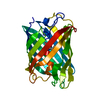 7qloC  5oqeS S: Starting model for refinement C: citing same article ( |
|---|---|
| Similar structure data |
-Links
 PDBj
PDBj

- Assembly
| Deposited unit | 
| ||||||||
|---|---|---|---|---|---|---|---|---|---|
| 1 |
| ||||||||
| Unit cell |
|
-Components
| #1: Protein | Mass: 25152.490 Da / Num. of mol.: 1 Source method: isolated from a genetically manipulated source Source: (gene. exp.) Lobophyllia hemprichii (invertebrata) / Production host:  |
|---|---|
| #2: Water | ChemComp-HOH /  Mass: 18.015 Da / Num. of mol.: 160 / Source method: isolated from a natural source / Formula: H2O Mass: 18.015 Da / Num. of mol.: 160 / Source method: isolated from a natural source / Formula: H2O |
| Has ligand of interest | Y |
-Experimental details
-Experiment
| Experiment | Method: X-RAY DIFFRACTION / Number of used crystals: 1 |
|---|
- Sample preparation
| Crystal | Density Matthews: 2.3 Å3/Da / Density % sol: 46.42 % |
|---|---|
| Crystal grow | Temperature: 293 K / Method: batch mode Details: 0.2 M LITHIUM SULFATE 0.1 M TRIS-CL PH 8.5 23-30% PEG 3350 |
-Data collection
| Diffraction | Mean temperature: 293 K / Serial crystal experiment: Y |
|---|---|
| Diffraction source | Source: FREE ELECTRON LASER / Site: SLAC LCLS  / Beamline: CXI / Wavelength: 1.305 Å / Beamline: CXI / Wavelength: 1.305 Å |
| Detector | Type: CS-PAD CXI-1 / Detector: PIXEL / Date: Feb 2, 2018 |
| Radiation | Protocol: SINGLE WAVELENGTH / Monochromatic (M) / Laue (L): M / Scattering type: x-ray |
| Radiation wavelength | Wavelength: 1.305 Å / Relative weight: 1 |
| Reflection | Resolution: 1.3→15.4 Å / Num. obs: 54155 / % possible obs: 100 % / Redundancy: 273 % / Biso Wilson estimate: 35.68 Å2 / CC1/2: 0.99 / CC star: 1 / R split: 0.0968 / Net I/σ(I): 4.843 |
| Reflection shell | Resolution: 1.35→1.398 Å / Mean I/σ(I) obs: 0.4 / Num. unique obs: 647523 / R split: 2.91 |
| Serial crystallography measurement | Focal spot size: 1 µm2 / Pulse duration: 40 fsec. / Pulse photon energy: 9.5 keV / XFEL pulse repetition rate: 120 Hz |
| Serial crystallography sample delivery | Method: injection |
- Processing
| Software |
| ||||||||||||||||||||||||
|---|---|---|---|---|---|---|---|---|---|---|---|---|---|---|---|---|---|---|---|---|---|---|---|---|---|
| Refinement | Method to determine structure: MOLECULAR REPLACEMENT Starting model: 5OQE Resolution: 1.3→15.4 Å / Cor.coef. Fo:Fc: 0.966 / Cor.coef. Fo:Fc free: 0.968 / SU B: 0.003 / SU ML: 0 / Cross valid method: THROUGHOUT / σ(F): 0 / ESU R: 0.051 / ESU R Free: 0.05 / Stereochemistry target values: MAXIMUM LIKELIHOOD Details: HYDROGENS HAVE BEEN ADDED IN THE RIDING POSITIONS U VALUES : REFINED INDIVIDUALLY
| ||||||||||||||||||||||||
| Solvent computation | Ion probe radii: 0.8 Å / Shrinkage radii: 0.8 Å / VDW probe radii: 1.2 Å / Solvent model: MASK | ||||||||||||||||||||||||
| Displacement parameters | Biso max: 69.13 Å2 / Biso mean: 16.918 Å2 / Biso min: 7.71 Å2
| ||||||||||||||||||||||||
| Refinement step | Cycle: final / Resolution: 1.3→15.4 Å
| ||||||||||||||||||||||||
| LS refinement shell | Resolution: 1.3→1.334 Å / Rfactor Rfree error: 0 / Total num. of bins used: 20
|
