 Movie
Movie Controller

[English] 日本語
 Yorodumi
- PDB-7qjl: Crystal structure of CYP142 from Mycobacterium tuberculosis in co... -
+ Open data

- Basic information
| Entry | Database: PDB / ID: 7qjl | ||||||
|---|---|---|---|---|---|---|---|
| Title | Crystal structure of CYP142 from Mycobacterium tuberculosis in complex with an inhibitor | ||||||
 Components Components | Steroid C26-monooxygenase | ||||||
 Keywords Keywords | OXIDOREDUCTASE / CYP / P450 / Complex / Inhibitor / TB / Tuberculosis / cholesterol / Cytochrome / Heme | ||||||
| Function / homology |  Function and homology information Function and homology informationcholesterol 26-hydroxylase activity / cholest-4-en-3-one 26-monooxygenase [(25R)-3-oxocholest-4-en-26-oate forming] / cholest-4-en-3-one 26-monooxygenase activity / steroid hydroxylase activity / cholesterol catabolic process / peptidoglycan-based cell wall / iron ion binding / heme binding Similarity search - Function | ||||||
| Biological species |   Mycobacterium tuberculosis (bacteria) Mycobacterium tuberculosis (bacteria) | ||||||
| Method |  X-RAY DIFFRACTION / SYNCHROTRON / MOLECULAR REPLACEMENT / Resolution: 1.38 Å X-RAY DIFFRACTION / SYNCHROTRON / MOLECULAR REPLACEMENT / Resolution: 1.38 Å | ||||||
 Authors Authors | Snee, M. / Levy, C. / Katariya, M. | ||||||
| Funding support |  United Kingdom, 1items United Kingdom, 1items
| ||||||
 Citation Citation | Journal: Chemistry / Year: 2023 Title: Structure Based Discovery of Inhibitors of CYP125 and CYP142 from Mycobacterium tuberculosis. Authors: Katariya, M.M. / Snee, M. / Tunnicliffe, R.B. / Kavanagh, M.E. / Boshoff, H.I.M. / Amadi, C.N. / Levy, C.W. / Munro, A.W. / Abell, C. / Leys, D. / Coyne, A.G. / McLean, K.J. | ||||||
| History |
|
- Structure visualization
| Structure viewer | Molecule: MolmilJmol/JSmol |
|---|
- Downloads & links
-Download
| PDBx/mmCIF format | 7qjl.cif.gz | 324.8 KB | Display | PDBx/mmCIF format |
|---|---|---|---|---|
| PDB format | pdb7qjl.ent.gz | 222.9 KB | Display | PDB format |
| PDBx/mmJSON format | 7qjl.json.gz | Tree view | PDBx/mmJSON format | |
| Others |  Other downloads Other downloads |
-Validation report
| Arichive directory | https://data.pdbj.org/pub/pdb/validation_reports/qj/7qjlftp://data.pdbj.org/pub/pdb/validation_reports/qj/7qjl | HTTPS FTP |
|---|
-Related structure data
| Related structure data |  7qkeC 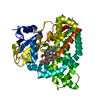 7qnnC  7qwnC  7r1iC 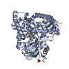 7r3uC  7yxfC  7zlzC  7zqrC  7zsuC  7zt0C  7zxdC 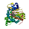 2xkrS C: citing same article ( S: Starting model for refinement |
|---|---|
| Similar structure data |
-Links
 PDBj
PDBj


- Assembly
| Deposited unit | 
| ||||||||||
|---|---|---|---|---|---|---|---|---|---|---|---|
| 1 |
| ||||||||||
| Unit cell |
|
-Components
-Protein , 1 types, 1 molecules A
| #1: Protein | Mass: 44371.238 Da / Num. of mol.: 1 Source method: isolated from a genetically manipulated source Source: (gene. exp.) Mycobacterium tuberculosis (bacteria) / Gene: cyp142, cyp142A1, Rv3518c, MTV023.25c / Plasmid: Pet21a / Production host: References: UniProt: P9WPL5, cholest-4-en-3-one 26-monooxygenase [(25R)-3-oxocholest-4-en-26-oate forming] |
|---|
-Non-polymers , 5 types, 499 molecules 


| #2: Chemical | ChemComp-HEM /  Mass: 616.487 Da / Num. of mol.: 1 / Source method: obtained synthetically / Formula: C34H32FeN4O4 Mass: 616.487 Da / Num. of mol.: 1 / Source method: obtained synthetically / Formula: C34H32FeN4O4 | ||||
|---|---|---|---|---|---|
| #3: Chemical | ChemComp-ACT /  Mass: 59.044 Da / Num. of mol.: 1 / Source method: obtained synthetically / Formula: C2H3O2 Mass: 59.044 Da / Num. of mol.: 1 / Source method: obtained synthetically / Formula: C2H3O2 | ||||
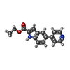
| #4: Chemical | ChemComp-BR /  Mass: 79.904 Da / Num. of mol.: 4 / Source method: obtained synthetically / Formula: Br Mass: 79.904 Da / Num. of mol.: 4 / Source method: obtained synthetically / Formula: Br#5: Chemical | ChemComp-DQE / |  Mass: 266.295 Da / Num. of mol.: 1 / Source method: obtained synthetically / Formula: C16H14N2O2 / Feature type: SUBJECT OF INVESTIGATION Mass: 266.295 Da / Num. of mol.: 1 / Source method: obtained synthetically / Formula: C16H14N2O2 / Feature type: SUBJECT OF INVESTIGATION#6: Water | ChemComp-HOH / | Mass: 18.015 Da / Num. of mol.: 492 / Source method: isolated from a natural source / Formula: H2O |
-Details
| Has ligand of interest | Y |
|---|
-Experimental details
-Experiment
| Experiment | Method: X-RAY DIFFRACTION / Number of used crystals: 1 |
|---|
- Sample preparation
| Crystal | Density Matthews: 2.69 Å3/Da / Density % sol: 54.3 % |
|---|---|
| Crystal grow | Temperature: 277.15 K / Method: vapor diffusion, sitting drop / pH: 4.5 Details: 8% w/v PEG 20,000, 8% v/v PEG 550 MME, 0.1M Sodium acetate pH 4.5, 0.25M KBr Temp details: 4C |
-Data collection
| Diffraction | Mean temperature: 100 K / Ambient temp details: N2 cryostream / Serial crystal experiment: N |
|---|---|
| Diffraction source | Source: SYNCHROTRON / Site: Diamond / Beamline: I03 / Wavelength: 0.9763 Å |
| Detector | Type: DECTRIS PILATUS3 6M / Detector: PIXEL / Date: Nov 25, 2018 |
| Radiation | Protocol: SINGLE WAVELENGTH / Monochromatic (M) / Laue (L): M / Scattering type: x-ray |
| Radiation wavelength | Wavelength: 0.9763 Å / Relative weight: 1 |
| Reflection | Resolution: 1.38→66.17 Å / Num. obs: 99205 / % possible obs: 100 % / Redundancy: 6.9 % / Biso Wilson estimate: 18 Å2 / CC1/2: 0.996 / Rmerge(I) obs: 0.087 / Rpim(I) all: 0.035 / Net I/σ(I): 8.2 |
| Reflection shell | Resolution: 1.38→1.4 Å / Redundancy: 6.4 % / Rmerge(I) obs: 1.509 / Mean I/σ(I) obs: 1.1 / Num. unique obs: 4848 / CC1/2: 0.527 / Rpim(I) all: 0.636 / % possible all: 100 |
- Processing
| Software |
| |||||||||||||||||||||||||||||||||||||||||||||||||||||||||||||||||||||||||||||||||||||||||||||||||||||||||||||||||||||||||||||||||||||||||||||||||||||||||||||||||||||||||||||||||||||||||||||||||||||||||||||||||||||||||
|---|---|---|---|---|---|---|---|---|---|---|---|---|---|---|---|---|---|---|---|---|---|---|---|---|---|---|---|---|---|---|---|---|---|---|---|---|---|---|---|---|---|---|---|---|---|---|---|---|---|---|---|---|---|---|---|---|---|---|---|---|---|---|---|---|---|---|---|---|---|---|---|---|---|---|---|---|---|---|---|---|---|---|---|---|---|---|---|---|---|---|---|---|---|---|---|---|---|---|---|---|---|---|---|---|---|---|---|---|---|---|---|---|---|---|---|---|---|---|---|---|---|---|---|---|---|---|---|---|---|---|---|---|---|---|---|---|---|---|---|---|---|---|---|---|---|---|---|---|---|---|---|---|---|---|---|---|---|---|---|---|---|---|---|---|---|---|---|---|---|---|---|---|---|---|---|---|---|---|---|---|---|---|---|---|---|---|---|---|---|---|---|---|---|---|---|---|---|---|---|---|---|---|---|---|---|---|---|---|---|---|---|---|---|---|---|---|---|---|
| Refinement | Method to determine structure: MOLECULAR REPLACEMENT Starting model: 2XKR Resolution: 1.38→51.06 Å / SU ML: 0.1346 / Cross valid method: FREE R-VALUE / σ(F): 1.33 / Phase error: 15.6389 Stereochemistry target values: GeoStd + Monomer Library + CDL v1.2
| |||||||||||||||||||||||||||||||||||||||||||||||||||||||||||||||||||||||||||||||||||||||||||||||||||||||||||||||||||||||||||||||||||||||||||||||||||||||||||||||||||||||||||||||||||||||||||||||||||||||||||||||||||||||||
| Solvent computation | Shrinkage radii: 0.9 Å / VDW probe radii: 1.11 Å / Solvent model: FLAT BULK SOLVENT MODEL | |||||||||||||||||||||||||||||||||||||||||||||||||||||||||||||||||||||||||||||||||||||||||||||||||||||||||||||||||||||||||||||||||||||||||||||||||||||||||||||||||||||||||||||||||||||||||||||||||||||||||||||||||||||||||
| Displacement parameters | Biso mean: 24.85 Å2 | |||||||||||||||||||||||||||||||||||||||||||||||||||||||||||||||||||||||||||||||||||||||||||||||||||||||||||||||||||||||||||||||||||||||||||||||||||||||||||||||||||||||||||||||||||||||||||||||||||||||||||||||||||||||||
| Refinement step | Cycle: LAST / Resolution: 1.38→51.06 Å
| |||||||||||||||||||||||||||||||||||||||||||||||||||||||||||||||||||||||||||||||||||||||||||||||||||||||||||||||||||||||||||||||||||||||||||||||||||||||||||||||||||||||||||||||||||||||||||||||||||||||||||||||||||||||||
| Refine LS restraints |
| |||||||||||||||||||||||||||||||||||||||||||||||||||||||||||||||||||||||||||||||||||||||||||||||||||||||||||||||||||||||||||||||||||||||||||||||||||||||||||||||||||||||||||||||||||||||||||||||||||||||||||||||||||||||||
| LS refinement shell |
| |||||||||||||||||||||||||||||||||||||||||||||||||||||||||||||||||||||||||||||||||||||||||||||||||||||||||||||||||||||||||||||||||||||||||||||||||||||||||||||||||||||||||||||||||||||||||||||||||||||||||||||||||||||||||
| Refinement TLS params. | Method: refined / Origin x: 15.3270247399 Å / Origin y: 45.1534219368 Å / Origin z: 82.6401864379 Å
| |||||||||||||||||||||||||||||||||||||||||||||||||||||||||||||||||||||||||||||||||||||||||||||||||||||||||||||||||||||||||||||||||||||||||||||||||||||||||||||||||||||||||||||||||||||||||||||||||||||||||||||||||||||||||
| Refinement TLS group | Selection details: all |
