 Movie
Movie Controller

[English] 日本語
 Yorodumi
- PDB-7qh8: CRYSTAL STRUCTURE OF LYSYL-TRNA SYNTHETASE FROM Mycobacterium tub... -
+ Open data

- Basic information
| Entry | Database: PDB / ID: 7qh8 | ||||||
|---|---|---|---|---|---|---|---|
| Title | CRYSTAL STRUCTURE OF LYSYL-TRNA SYNTHETASE FROM Mycobacterium tuberculosis COMPLEXED WITH L-LYSINE | ||||||
 Components Components | Lysine--tRNA ligase 1 | ||||||
 Keywords Keywords | LIGASE / Mycobacterium tuberculosis / ATP binding | ||||||
| Function / homology |  Function and homology information Function and homology informationlysine-tRNA ligase / lysine-tRNA ligase activity / lysyl-tRNA aminoacylation / tRNA binding / magnesium ion binding / ATP binding / cytoplasm / cytosol Similarity search - Function | ||||||
| Biological species |  Mycobacterium tuberculosis H37Rv (bacteria) Mycobacterium tuberculosis H37Rv (bacteria) | ||||||
| Method |  X-RAY DIFFRACTION / SYNCHROTRON / MOLECULAR REPLACEMENT / Resolution: 1.92 Å X-RAY DIFFRACTION / SYNCHROTRON / MOLECULAR REPLACEMENT / Resolution: 1.92 Å | ||||||
 Authors Authors | Dawson, A. / Robinson, D.A. / Tamjar, J. / Wyatt, P. / Green, S. | ||||||
| Funding support |  United States, 1items United States, 1items
| ||||||
 Citation Citation | Journal: Nat Commun / Year: 2022 Title: Lysyl-tRNA synthetase, a target for urgently needed M. tuberculosis drugs. Authors: Green, S.R. / Davis, S.H. / Damerow, S. / Engelhart, C.A. / Mathieson, M. / Baragana, B. / Robinson, D.A. / Tamjar, J. / Dawson, A. / Tamaki, F.K. / Buchanan, K.I. / Post, J. / Dowers, K. / ...Authors: Green, S.R. / Davis, S.H. / Damerow, S. / Engelhart, C.A. / Mathieson, M. / Baragana, B. / Robinson, D.A. / Tamjar, J. / Dawson, A. / Tamaki, F.K. / Buchanan, K.I. / Post, J. / Dowers, K. / Shepherd, S.M. / Jansen, C. / Zuccotto, F. / Gilbert, I.H. / Epemolu, O. / Riley, J. / Stojanovski, L. / Osuna-Cabello, M. / Perez-Herran, E. / Rebollo, M.J. / Guijarro Lopez, L. / Casado Castro, P. / Camino, I. / Kim, H.C. / Bean, J.M. / Nahiyaan, N. / Rhee, K.Y. / Wang, Q. / Tan, V.Y. / Boshoff, H.I.M. / Converse, P.J. / Li, S.Y. / Chang, Y.S. / Fotouhi, N. / Upton, A.M. / Nuermberger, E.L. / Schnappinger, D. / Read, K.D. / Encinas, L. / Bates, R.H. / Wyatt, P.G. / Cleghorn, L.A.T. | ||||||
| History |
|
- Structure visualization
| Structure viewer | Molecule: MolmilJmol/JSmol |
|---|
- Downloads & links
-Download
| PDBx/mmCIF format | 7qh8.cif.gz | 110.6 KB | Display | PDBx/mmCIF format |
|---|---|---|---|---|
| PDB format | pdb7qh8.ent.gz | 81.7 KB | Display | PDB format |
| PDBx/mmJSON format | 7qh8.json.gz | Tree view | PDBx/mmJSON format | |
| Others |  Other downloads Other downloads |
-Validation report
| Arichive directory | https://data.pdbj.org/pub/pdb/validation_reports/qh/7qh8ftp://data.pdbj.org/pub/pdb/validation_reports/qh/7qh8 | HTTPS FTP |
|---|
-Related structure data
| Related structure data |  7qhnC  7qi8C  5eloS S: Starting model for refinement C: citing same article ( |
|---|---|
| Similar structure data |
-Links
 PDBj
PDBj

- Assembly
| Deposited unit | 
| ||||||||
|---|---|---|---|---|---|---|---|---|---|
| 1 | 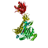
| ||||||||
| Unit cell |
|
-Components
| #1: Protein | Mass: 58189.691 Da / Num. of mol.: 1 Source method: isolated from a genetically manipulated source Source: (gene. exp.) Mycobacterium tuberculosis H37Rv (bacteria)Gene: lysS1, lysS, Rv3598c, MTCY07H7B.24 / Production host: |
|---|---|
| #2: Chemical | ChemComp-LYS /   Type: L-peptide linking / Mass: 147.195 Da / Num. of mol.: 1 / Source method: obtained synthetically / Formula: C6H15N2O2 Type: L-peptide linking / Mass: 147.195 Da / Num. of mol.: 1 / Source method: obtained synthetically / Formula: C6H15N2O2 |
| #3: Chemical | ChemComp-GOL /   Mass: 92.094 Da / Num. of mol.: 1 / Source method: isolated from a natural source / Formula: C3H8O3 Mass: 92.094 Da / Num. of mol.: 1 / Source method: isolated from a natural source / Formula: C3H8O3 |
| #4: Water | ChemComp-HOH /  Mass: 18.015 Da / Num. of mol.: 131 / Source method: isolated from a natural source / Formula: H2O Mass: 18.015 Da / Num. of mol.: 131 / Source method: isolated from a natural source / Formula: H2O |
| Has ligand of interest | N |
-Experimental details
-Experiment
| Experiment | Method: X-RAY DIFFRACTION / Number of used crystals: 1 |
|---|
- Sample preparation
| Crystal | Density Matthews: 2.23 Å3/Da / Density % sol: 44.88 % / Description: block |
|---|---|
| Crystal grow | Temperature: 293 K / Method: vapor diffusion, hanging drop / pH: 7 Details: Reservoir: 0.25 M NaOAc, 14% w/v PEG 3350 Protein buffer: 25 mM HEPES, 500 mM NaCl, 5% glycerol, 2 mM DTT, pH 7.0 Temp details: room temperature |
-Data collection
| Diffraction | Mean temperature: 100 K / Serial crystal experiment: N |
|---|---|
| Diffraction source | Source: SYNCHROTRON / Site: Diamond  / Beamline: I04 / Wavelength: 0.9795 Å / Beamline: I04 / Wavelength: 0.9795 Å |
| Detector | Type: DECTRIS PILATUS 6M-F / Detector: PIXEL / Date: Sep 9, 2018 |
| Radiation | Protocol: SINGLE WAVELENGTH / Monochromatic (M) / Laue (L): M / Scattering type: x-ray |
| Radiation wavelength | Wavelength: 0.9795 Å / Relative weight: 1 |
| Reflection | Resolution: 1.92→73.89 Å / Num. obs: 41053 / % possible obs: 100 % / Redundancy: 13.8 % / CC1/2: 0.999 / Rmerge(I) obs: 0.095 / Rpim(I) all: 0.026 / Rrim(I) all: 0.099 / Net I/σ(I): 15.1 |
| Reflection shell | Resolution: 1.92→1.95 Å / Redundancy: 12 % / Rmerge(I) obs: 3.304 / Mean I/σ(I) obs: 1 / Num. unique obs: 2007 / CC1/2: 0.731 / Rpim(I) all: 0.992 / % possible all: 99.6 |
- Processing
| Software |
| ||||||||||||||||||||||||||||||||||||||||||||||||||||||||||||
|---|---|---|---|---|---|---|---|---|---|---|---|---|---|---|---|---|---|---|---|---|---|---|---|---|---|---|---|---|---|---|---|---|---|---|---|---|---|---|---|---|---|---|---|---|---|---|---|---|---|---|---|---|---|---|---|---|---|---|---|---|---|
| Refinement | Method to determine structure: MOLECULAR REPLACEMENT Starting model: 5elo Resolution: 1.92→59.35 Å / Cor.coef. Fo:Fc: 0.962 / Cor.coef. Fo:Fc free: 0.95 / SU B: 5.536 / SU ML: 0.149 / Cross valid method: THROUGHOUT / σ(F): 0 / ESU R: 0.165 / ESU R Free: 0.151 / Stereochemistry target values: MAXIMUM LIKELIHOOD Details: HYDROGENS HAVE BEEN ADDED IN THE RIDING POSITIONS U VALUES : REFINED INDIVIDUALLY
| ||||||||||||||||||||||||||||||||||||||||||||||||||||||||||||
| Solvent computation | Ion probe radii: 0.8 Å / Shrinkage radii: 0.8 Å / VDW probe radii: 1.2 Å / Solvent model: MASK | ||||||||||||||||||||||||||||||||||||||||||||||||||||||||||||
| Displacement parameters | Biso max: 124.9 Å2 / Biso mean: 49.157 Å2 / Biso min: 27.71 Å2
| ||||||||||||||||||||||||||||||||||||||||||||||||||||||||||||
| Refinement step | Cycle: final / Resolution: 1.92→59.35 Å
| ||||||||||||||||||||||||||||||||||||||||||||||||||||||||||||
| Refine LS restraints |
| ||||||||||||||||||||||||||||||||||||||||||||||||||||||||||||
| LS refinement shell | Resolution: 1.92→1.97 Å / Rfactor Rfree error: 0 / Total num. of bins used: 20
|
