 Movie
Movie Controller

[English] 日本語
 Yorodumi
- PDB-7qfx: Crystal structure of Old Yellow Enzyme AnOYE8 from Aspergillus niger -
+ Open data

- Basic information
| Entry | Database: PDB / ID: 7qfx | ||||||
|---|---|---|---|---|---|---|---|
| Title | Crystal structure of Old Yellow Enzyme AnOYE8 from Aspergillus niger | ||||||
 Components Components | NADH-dependent flavin oxidoreductase | ||||||
 Keywords Keywords | OXIDOREDUCTASE / Old Yellow Enzyme / Ene-reductase | ||||||
| Function / homology |  Function and homology information Function and homology information | ||||||
| Biological species |  | ||||||
| Method |  X-RAY DIFFRACTION / SYNCHROTRON / MOLECULAR REPLACEMENT / Resolution: 2.8 Å X-RAY DIFFRACTION / SYNCHROTRON / MOLECULAR REPLACEMENT / Resolution: 2.8 Å | ||||||
 Authors Authors | Robescu, M.S. / Loprete, G. / Vascon, F. / Gasparotto, M. / Filippini, F. / Bergantino, E. / Cendron, L. | ||||||
| Funding support | 1items
| ||||||
 Citation Citation | Journal: Int J Mol Sci / Year: 2022 Title: The Family Keeps on Growing: Four Novel Fungal OYEs Characterized. Authors: Robescu, M.S. / Loprete, G. / Gasparotto, M. / Vascon, F. / Filippini, F. / Cendron, L. / Bergantino, E. #1: Journal: Acta Crystallogr., Sect. D: Biol. Crystallogr. / Year: 2012Title: Towards automated crystallographic structure refinement with phenix.refine. Authors: Afonine, P.V. | ||||||
| History |
|
- Structure visualization
| Structure viewer | Molecule: MolmilJmol/JSmol |
|---|
- Downloads & links
-Download
| PDBx/mmCIF format | 7qfx.cif.gz | 637.3 KB | Display | PDBx/mmCIF format |
|---|---|---|---|---|
| PDB format | pdb7qfx.ent.gz | 529.8 KB | Display | PDB format |
| PDBx/mmJSON format | 7qfx.json.gz | Tree view | PDBx/mmJSON format | |
| Others |  Other downloads Other downloads |
-Validation report
| Summary document | 7qfx_validation.pdf.gz | 1.7 MB | Display | wwPDB validaton report |
|---|---|---|---|---|
| Full document | 7qfx_full_validation.pdf.gz | 1.8 MB | Display | |
| Data in XML | 7qfx_validation.xml.gz | 65.2 KB | Display | |
| Data in CIF | 7qfx_validation.cif.gz | 86.4 KB | Display | |
| Arichive directory | https://data.pdbj.org/pub/pdb/validation_reports/qf/7qfxftp://data.pdbj.org/pub/pdb/validation_reports/qf/7qfx | HTTPS FTP |
-Related structure data
| Related structure data |  5lnjS S: Starting model for refinement |
|---|---|
| Similar structure data |
-Links
 PDBj
PDBj- Assembly
| Deposited unit | 
| ||||||||||
|---|---|---|---|---|---|---|---|---|---|---|---|
| 1 | 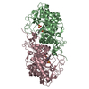
| ||||||||||
| 2 | 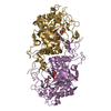
| ||||||||||
| Unit cell |
|
-Components
| #1: Protein | Mass: 45910.000 Da / Num. of mol.: 4 Source method: isolated from a genetically manipulated source Source: (gene. exp.)  #2: Chemical | ChemComp-SO4 /   Mass: 96.063 Da / Num. of mol.: 4 / Source method: obtained synthetically / Formula: SO4 Mass: 96.063 Da / Num. of mol.: 4 / Source method: obtained synthetically / Formula: SO4#3: Chemical | ChemComp-FMN /   Mass: 456.344 Da / Num. of mol.: 4 / Source method: obtained synthetically / Formula: C17H21N4O9P / Feature type: SUBJECT OF INVESTIGATION Mass: 456.344 Da / Num. of mol.: 4 / Source method: obtained synthetically / Formula: C17H21N4O9P / Feature type: SUBJECT OF INVESTIGATION#4: Chemical | ChemComp-MPD / ( |   Mass: 118.174 Da / Num. of mol.: 1 / Source method: obtained synthetically / Formula: C6H14O2 / Comment: precipitant*YM Mass: 118.174 Da / Num. of mol.: 1 / Source method: obtained synthetically / Formula: C6H14O2 / Comment: precipitant*YM#5: Water | ChemComp-HOH / |  Mass: 18.015 Da / Num. of mol.: 85 / Source method: isolated from a natural source / Formula: H2O Mass: 18.015 Da / Num. of mol.: 85 / Source method: isolated from a natural source / Formula: H2OHas ligand of interest | Y | |
|---|
-Experimental details
-Experiment
| Experiment | Method: X-RAY DIFFRACTION / Number of used crystals: 1 |
|---|
- Sample preparation
| Crystal | Density Matthews: 2.43 Å3/Da / Density % sol: 49.34 % |
|---|---|
| Crystal grow | Temperature: 293 K / Method: vapor diffusion, sitting drop Details: 18 % w/v PEG 5000 MME, 0.1 M MES pH 6.5, 0.2 M ammonium sulphate |
-Data collection
| Diffraction | Mean temperature: 100 K / Serial crystal experiment: N |
|---|---|
| Diffraction source | Source: SYNCHROTRON / Site: ESRF  / Beamline: ID23-2 / Wavelength: 0.873 Å / Beamline: ID23-2 / Wavelength: 0.873 Å |
| Detector | Type: DECTRIS PILATUS 6M / Detector: PIXEL / Date: Jan 7, 2021 |
| Radiation | Protocol: SINGLE WAVELENGTH / Monochromatic (M) / Laue (L): M / Scattering type: x-ray |
| Radiation wavelength | Wavelength: 0.873 Å / Relative weight: 1 |
| Reflection | Resolution: 2.8→39.98 Å / Num. obs: 43316 / % possible obs: 97 % / Redundancy: 4.8 % / Biso Wilson estimate: 52.61 Å2 / CC1/2: 0.992 / Rmerge(I) obs: 0.131 / Net I/σ(I): 13.2 |
| Reflection shell | Resolution: 2.8→2.89 Å / Redundancy: 4.2 % / Rmerge(I) obs: 0.595 / Mean I/σ(I) obs: 3 / Num. unique obs: 4327 / % possible all: 95 |
- Processing
| Software |
| ||||||||||||||||||||
|---|---|---|---|---|---|---|---|---|---|---|---|---|---|---|---|---|---|---|---|---|---|
| Refinement | Method to determine structure: MOLECULAR REPLACEMENT Starting model: 5LNJ Resolution: 2.8→39.95 Å / Cross valid method: THROUGHOUT
| ||||||||||||||||||||
| Displacement parameters | Biso max: 151.91 Å2 / Biso mean: 72.6487 Å2 / Biso min: 30 Å2 | ||||||||||||||||||||
| Refinement step | Cycle: LAST / Resolution: 2.8→39.95 Å
|
