 Movie
Movie Controller

[English] 日本語
 Yorodumi
- PDB-7qar: Serial crystallography structure of cofactor-free urate oxidase i... -
+ Open data

- Basic information
| Entry | Database: PDB / ID: 7qar | ||||||
|---|---|---|---|---|---|---|---|
| Title | Serial crystallography structure of cofactor-free urate oxidase in complex with the 5-peroxo derivative of 9-methyl uric acid at room temperature | ||||||
 Components Components | Uricase | ||||||
 Keywords Keywords | OXIDOREDUCTASE / COFACTOR-FREE OXIDASE / REACTION INTERMEDIATE / SERIAL CRYSTALLOGRAPHY / DIOXYGEN | ||||||
| Function / homology |  Function and homology information Function and homology informationfactor-independent urate hydroxylase / urate oxidase activity / purine nucleobase catabolic process / urate catabolic process / peroxisome Similarity search - Function | ||||||
| Biological species |  | ||||||
| Method |  X-RAY DIFFRACTION / SYNCHROTRON / MOLECULAR REPLACEMENT / Resolution: 2.3 Å X-RAY DIFFRACTION / SYNCHROTRON / MOLECULAR REPLACEMENT / Resolution: 2.3 Å | ||||||
 Authors Authors | Bui, S. / Catapano, L. / Zielinski, K. / Yefanov, O. / Murshudov, G.N. / Oberthuer, D. / Steiner, R.A. | ||||||
| Funding support |  United Kingdom, 1items United Kingdom, 1items
| ||||||
 Citation Citation | Journal: Iucrj / Year: 2022 Title: Rapid and efficient room-temperature serial synchrotron crystallography using the CFEL TapeDrive. Authors: Zielinski, K.A. / Prester, A. / Andaleeb, H. / Bui, S. / Yefanov, O. / Catapano, L. / Henkel, A. / Wiedorn, M.O. / Lorbeer, O. / Crosas, E. / Meyer, J. / Mariani, V. / Domaracky, M. / White, ...Authors: Zielinski, K.A. / Prester, A. / Andaleeb, H. / Bui, S. / Yefanov, O. / Catapano, L. / Henkel, A. / Wiedorn, M.O. / Lorbeer, O. / Crosas, E. / Meyer, J. / Mariani, V. / Domaracky, M. / White, T.A. / Fleckenstein, H. / Sarrou, I. / Werner, N. / Betzel, C. / Rohde, H. / Aepfelbacher, M. / Chapman, H.N. / Perbandt, M. / Steiner, R.A. / Oberthuer, D. | ||||||
| History |
|
- Structure visualization
| Structure viewer | Molecule: MolmilJmol/JSmol |
|---|
- Downloads & links
-Download
| PDBx/mmCIF format | 7qar.cif.gz | 80.2 KB | Display | PDBx/mmCIF format |
|---|---|---|---|---|
| PDB format | pdb7qar.ent.gz | Display | PDB format | |
| PDBx/mmJSON format | 7qar.json.gz | Tree view | PDBx/mmJSON format | |
| Others |  Other downloads Other downloads |
-Validation report
| Arichive directory | https://data.pdbj.org/pub/pdb/validation_reports/qa/7qarftp://data.pdbj.org/pub/pdb/validation_reports/qa/7qar | HTTPS FTP |
|---|
-Related structure data
| Related structure data | 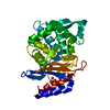 7zpvC 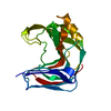 7zq0C  8af4C 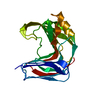 8af5C  8af6C 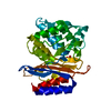 8af7C 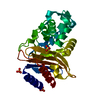 8af8C  4cw2S S: Starting model for refinement C: citing same article ( |
|---|---|
| Similar structure data |
-Links
 PDBj
PDBj
- Assembly
| Deposited unit | 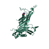
| |||||||||
|---|---|---|---|---|---|---|---|---|---|---|
| 1 | 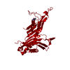
| |||||||||
| Unit cell |
| |||||||||
| Components on special symmetry positions |
|
-Components
| #1: Protein | Mass: 34288.742 Da / Num. of mol.: 1 Source method: isolated from a genetically manipulated source Source: (gene. exp.)  References: UniProt: Q00511, factor-independent urate hydroxylase |
|---|---|
| #2: Chemical | ChemComp-XDS / (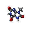  Mass: 214.136 Da / Num. of mol.: 1 / Source method: obtained synthetically / Formula: C6H6N4O5 / Feature type: SUBJECT OF INVESTIGATION Mass: 214.136 Da / Num. of mol.: 1 / Source method: obtained synthetically / Formula: C6H6N4O5 / Feature type: SUBJECT OF INVESTIGATION |
| #3: Water | ChemComp-HOH /  Mass: 18.015 Da / Num. of mol.: 127 / Source method: isolated from a natural source / Formula: H2O Mass: 18.015 Da / Num. of mol.: 127 / Source method: isolated from a natural source / Formula: H2O |
| Has ligand of interest | Y |
-Experimental details
-Experiment
| Experiment | Method: X-RAY DIFFRACTION / Number of used crystals: 1 |
|---|
- Sample preparation
| Crystal | Density Matthews: 3.01 Å3/Da / Density % sol: 59.11 % |
|---|---|
| Crystal grow | Temperature: 291 K / Method: batch mode / pH: 7.6 / Details: TRIS-ACETATE, PEG 8000, MUA, |
-Data collection
| Diffraction | Mean temperature: 293 K / Serial crystal experiment: Y |
|---|---|
| Diffraction source | Source: SYNCHROTRON / Site: PETRA III, DESY  / Beamline: P11 / Wavelength: 1.03 Å / Beamline: P11 / Wavelength: 1.03 Å |
| Detector | Type: DECTRIS PILATUS3 6M / Detector: PIXEL / Date: Apr 23, 2019 |
| Radiation | Protocol: SINGLE WAVELENGTH / Monochromatic (M) / Laue (L): M / Scattering type: x-ray |
| Radiation wavelength | Wavelength: 1.03 Å / Relative weight: 1 |
| Reflection | Resolution: 2.3→17.07 Å / Num. obs: 18280 / % possible obs: 99.98 % / Redundancy: 28.2 % / CC1/2: 0.849 / CC star: 0.958 / R split: 0.3668 / Net I/σ(I): 2.44 |
| Reflection shell | Resolution: 2.3→2.34 Å / Redundancy: 20.6 % / Mean I/σ(I) obs: 0.52 / Num. unique obs: 1796 / CC1/2: 0.225 / CC star: 0.606 / R split: 1.9369 / % possible all: 100 |
| Serial crystallography sample delivery | Method: injection |
- Processing
| Software |
| |||||||||||||||||||||||||||||||||||||||||||||||||||||||||||||||||||||||||||||||||||||||||||||||||||||||||||||||||||||||||||||||||||||||||||||||||||||||||||
|---|---|---|---|---|---|---|---|---|---|---|---|---|---|---|---|---|---|---|---|---|---|---|---|---|---|---|---|---|---|---|---|---|---|---|---|---|---|---|---|---|---|---|---|---|---|---|---|---|---|---|---|---|---|---|---|---|---|---|---|---|---|---|---|---|---|---|---|---|---|---|---|---|---|---|---|---|---|---|---|---|---|---|---|---|---|---|---|---|---|---|---|---|---|---|---|---|---|---|---|---|---|---|---|---|---|---|---|---|---|---|---|---|---|---|---|---|---|---|---|---|---|---|---|---|---|---|---|---|---|---|---|---|---|---|---|---|---|---|---|---|---|---|---|---|---|---|---|---|---|---|---|---|---|---|---|---|
| Refinement | Method to determine structure: MOLECULAR REPLACEMENT Starting model: 4CW2 Resolution: 2.3→17.07 Å / Cor.coef. Fo:Fc: 0.944 / Cor.coef. Fo:Fc free: 0.919 / SU B: 14.576 / SU ML: 0.297 / Cross valid method: FREE R-VALUE / ESU R: 0.309 / ESU R Free: 0.24 Details: Hydrogens have been added in their riding positions
| |||||||||||||||||||||||||||||||||||||||||||||||||||||||||||||||||||||||||||||||||||||||||||||||||||||||||||||||||||||||||||||||||||||||||||||||||||||||||||
| Solvent computation | Ion probe radii: 0.8 Å / Shrinkage radii: 0.8 Å / VDW probe radii: 1.2 Å / Solvent model: MASK BULK SOLVENT | |||||||||||||||||||||||||||||||||||||||||||||||||||||||||||||||||||||||||||||||||||||||||||||||||||||||||||||||||||||||||||||||||||||||||||||||||||||||||||
| Displacement parameters | Biso mean: 36.703 Å2
| |||||||||||||||||||||||||||||||||||||||||||||||||||||||||||||||||||||||||||||||||||||||||||||||||||||||||||||||||||||||||||||||||||||||||||||||||||||||||||
| Refinement step | Cycle: LAST / Resolution: 2.3→17.07 Å
| |||||||||||||||||||||||||||||||||||||||||||||||||||||||||||||||||||||||||||||||||||||||||||||||||||||||||||||||||||||||||||||||||||||||||||||||||||||||||||
| Refine LS restraints |
| |||||||||||||||||||||||||||||||||||||||||||||||||||||||||||||||||||||||||||||||||||||||||||||||||||||||||||||||||||||||||||||||||||||||||||||||||||||||||||
| LS refinement shell |
|
