| Entry | Database: PDB / ID: 7qac
|
|---|
| Title | The T2 structure of polycrystalline cubic human insulin |
|---|
 Components Components | - Insulin A chain
- Insulin B chain
|
|---|
 Keywords Keywords | HORMONE / cubic / human / insulin / T2 |
|---|
| Function / homology |  Function and homology information Function and homology information
negative regulation of glycogen catabolic process / positive regulation of nitric oxide mediated signal transduction / negative regulation of fatty acid metabolic process / negative regulation of feeding behavior / Signaling by Insulin receptor / IRS activation / regulation of protein secretion / Insulin processing / positive regulation of peptide hormone secretion / positive regulation of respiratory burst ...negative regulation of glycogen catabolic process / positive regulation of nitric oxide mediated signal transduction / negative regulation of fatty acid metabolic process / negative regulation of feeding behavior / Signaling by Insulin receptor / IRS activation / regulation of protein secretion / Insulin processing / positive regulation of peptide hormone secretion / positive regulation of respiratory burst / negative regulation of acute inflammatory response / Regulation of gene expression in beta cells / alpha-beta T cell activation / positive regulation of dendritic spine maintenance / Synthesis, secretion, and deacylation of Ghrelin / negative regulation of protein secretion / negative regulation of gluconeogenesis / positive regulation of glycogen biosynthetic process / fatty acid homeostasis / Signal attenuation / positive regulation of insulin receptor signaling pathway / negative regulation of respiratory burst involved in inflammatory response / FOXO-mediated transcription of oxidative stress, metabolic and neuronal genes / negative regulation of lipid catabolic process / positive regulation of lipid biosynthetic process / negative regulation of oxidative stress-induced intrinsic apoptotic signaling pathway / regulation of protein localization to plasma membrane / nitric oxide-cGMP-mediated signaling / transport vesicle / COPI-mediated anterograde transport / positive regulation of nitric-oxide synthase activity / Insulin receptor recycling / negative regulation of reactive oxygen species biosynthetic process / positive regulation of brown fat cell differentiation / insulin-like growth factor receptor binding / NPAS4 regulates expression of target genes / neuron projection maintenance / endoplasmic reticulum-Golgi intermediate compartment membrane / positive regulation of mitotic nuclear division / Insulin receptor signalling cascade / positive regulation of glycolytic process / positive regulation of cytokine production / endosome lumen / positive regulation of long-term synaptic potentiation / acute-phase response / positive regulation of D-glucose import across plasma membrane / positive regulation of protein secretion / insulin receptor binding / positive regulation of cell differentiation / Regulation of insulin secretion / wound healing / positive regulation of neuron projection development / hormone activity / regulation of synaptic plasticity / negative regulation of protein catabolic process / positive regulation of protein localization to nucleus / Golgi lumen / vasodilation / cognition / glucose metabolic process / insulin receptor signaling pathway / cell-cell signaling / glucose homeostasis / regulation of protein localization / PI5P, PP2A and IER3 Regulate PI3K/AKT Signaling / positive regulation of cell growth / protease binding / secretory granule lumen / positive regulation of canonical NF-kappaB signal transduction / positive regulation of phosphatidylinositol 3-kinase/protein kinase B signal transduction / positive regulation of MAPK cascade / positive regulation of cell migration / G protein-coupled receptor signaling pathway / endoplasmic reticulum lumen / Amyloid fiber formation / receptor ligand activity / Golgi membrane / negative regulation of gene expression / positive regulation of cell population proliferation / positive regulation of gene expression / regulation of DNA-templated transcription / extracellular space / extracellular region / identical protein bindingSimilarity search - Function Insulin / Insulin family / Insulin-like / Insulin/IGF/Relaxin family / Insulin / insulin-like growth factor / relaxin family. / Insulin, conserved site / Insulin family signature. / Insulin-like superfamilySimilarity search - Domain/homology |
|---|
| Biological species |  Homo sapiens (human) Homo sapiens (human) |
|---|
| Method | POWDER DIFFRACTION /  SYNCHROTRON / MOLECULAR REPLACEMENT / Resolution: 2.29 Å SYNCHROTRON / MOLECULAR REPLACEMENT / Resolution: 2.29 Å |
|---|
 Authors Authors | Karavassili, F. / Triandafillidis, D.P. / Valmas, A. / Spiliopoulou, M. / Fili, S. / Kontou, P. / Bowler, M.W. / Von Dreele, R.B. / Fitch, A. / Margiolaki, I. |
|---|
| Funding support | European Union,  Greece, 2items Greece, 2items | Organization | Grant number | Country |
|---|
| General Secretariat for Research and Technology (GSRT) | | European Union | | Hellenic Foundation for Research and Innovation (HFRI) | 3051 | Greece |
|
|---|
 Citation Citation | Journal: Acta Crystallogr D Struct Biol / Year: 2023
Title: The T 2 structure of polycrystalline cubic human insulin.
Authors: Triandafillidis, D.P. / Karavassili, F. / Spiliopoulou, M. / Valmas, A. / Athanasiadou, M. / Nikolaras, G. / Fili, S. / Kontou, P. / Bowler, M.W. / Chasapis, C.T. / Von Dreele, R.B. / Fitch, ...Authors: Triandafillidis, D.P. / Karavassili, F. / Spiliopoulou, M. / Valmas, A. / Athanasiadou, M. / Nikolaras, G. / Fili, S. / Kontou, P. / Bowler, M.W. / Chasapis, C.T. / Von Dreele, R.B. / Fitch, A.N. / Margiolaki, I. |
|---|
| History | | Deposition | Nov 16, 2021 | Deposition site: PDBE / Processing site: PDBE |
|---|
| Revision 1.0 | Jun 21, 2023 | Provider: repository / Type: Initial release |
|---|
| Revision 1.1 | Jul 19, 2023 | Group: Data collection / Category: diffrn_source / Item: _diffrn_source.type |
|---|
| Revision 1.2 | Aug 16, 2023 | Group: Data collection / Category: chem_comp_atom / chem_comp_bond |
|---|
| Revision 1.3 | Feb 7, 2024 | Group: Refinement description / Category: pdbx_initial_refinement_model / Item: _pdbx_initial_refinement_model.accession_code |
|---|
| Revision 1.4 | Oct 9, 2024 | Group: Structure summary / Category: pdbx_entry_details / pdbx_modification_feature |
|---|
|
|---|
| Remark 1 | REMARK 250 REFINEMENT. REMARK 250 PROGRAM : GSAS REMARK 250 AUTHORS : LARSON & VON DREELE REMARK ... REMARK 250 REFINEMENT. REMARK 250 PROGRAM : GSAS REMARK 250 AUTHORS : LARSON & VON DREELE REMARK 250 REMARK 250 DATA USED IN REFINEMENT REMARK 250 RESOLUTION RANGE HIGH (ANGSTROMS) : 2.29 REMARK 250 RESOLUTION RANGE LOW (ANGSTROMS) : 39.43 REMARK 250 POWDER DIFFRACTION DATA. REMARK 250 REMARK 250 FIT TO DATA USED IN REFINEMENT REMARK 250 NUMBER OF POWDER PATTERNS : 6 REMARK 250 PROFILE R VALUES (%) : 7.70 11.54 12.16 8.80 7.61 2.12 REMARK 250 WEIGHTED PROFILE R VALUES (%) : 10.98 14.75 15.17 12.12 10.95 3.34 REMARK 250 F**2 R VALUES (%) : 42.02 38.68 44.09 38.89 35.52 24.00 REMARK 250 NUMBERS OF POWDER PATTERN POINTS : 8256 7003 7002 7002 7002 4740 REMARK 250 NUMBERS OF REFLECTIONS : 3816 2345 2349 2345 2305 4998 REMARK 250 TOTAL NUMBER OF POWDER POINTS : 41005 REMARK 250 REMARK 250 NUMBER OF NON-HYDROGEN ATOMS USED IN REFINEMENT. REMARK 250 PROTEIN ATOMS : REMARK 250 NUCLEIC ACID ATOMS : REMARK 250 HETEROGEN ATOMS : REMARK 250 SOLVENT ATOMS : REMARK 250 REMARK 250 MODEL REFINEMENT. REMARK 250 NUMBER OF LEAST-SQUARES PARAMETERS : 786 REMARK 250 NUMBER OF RESTRAINTS : 1951 REMARK 250 LEAST-SQUARES MATRIX BAND WIDTH : 50 REMARK 250 MARQUARDT COEFFICIENT : 8.30 REMARK 250 REMARK 250 RMS DEVIATIONS FROM RESTRAINT TARGET VALUES. NUMBER. REMARK 250 INTERATOMIC DISTANCES (A) :0.015 506 REMARK 250 BOND ANGLES (DEG) : 1.43 702 REMARK 250 CHIRAL VOLUMES (A**3) :0.036 52 REMARK 250 TORSION ANGLE RESTRAINTS (E) : 1.00 98 REMARK 250 DISTANCES FROM RESTRAINT PLANES (A) :0.020 149 REMARK 250 TORSION PSEUDOPOTENTIAL RESTRAINTS (E) : 3.84 68 REMARK 250 ANTI-BUMPING DISTANCE RESTRAINTS (A) :0.123 186 REMARK 250 HYDROGEN BOND DISTANCE RESTRAINTS (A) :0.293 212 |
|---|
 Movie
Movie Controller


 Open data
 Basic information
 Structure visualization
 Downloads & links
 Other downloads
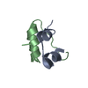
 PDBj
PDBj


 Assembly
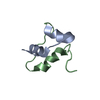

 Mass: 18.015 Da / Num. of mol.: 62 / Source method: isolated from a natural source / Formula: H2O
 Sample preparation