 Movie
Movie Controller

[English] 日本語
 Yorodumi
- PDB-7qa4: Crystal structure of stabilized H3N2 A/Hong Kong/1/1968 Hemagglut... -
+ Open data

- Basic information
| Entry | Database: PDB / ID: 7qa4 | ||||||
|---|---|---|---|---|---|---|---|
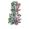
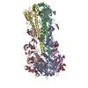
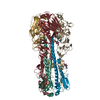
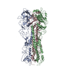
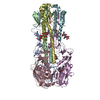
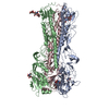
| Title | Crystal structure of stabilized H3N2 A/Hong Kong/1/1968 Hemagglutinin at 2.2 Angstrom | ||||||
 Components Components | Hemagglutinin | ||||||
 Keywords Keywords | VIRAL PROTEIN / Influenza / Hemagglutinin / Stabilized / Fusion protein / UNKNOWN FUNCTION | ||||||
| Function / homology |  Function and homology information Function and homology informationviral budding from plasma membrane / clathrin-dependent endocytosis of virus by host cell / host cell surface receptor binding / fusion of virus membrane with host plasma membrane / fusion of virus membrane with host endosome membrane / viral envelope / virion attachment to host cell / host cell plasma membrane / virion membrane / membrane Similarity search - Function | ||||||
| Biological species |   Influenza A virus Influenza A virus | ||||||
| Method |  X-RAY DIFFRACTION / SYNCHROTRON / MOLECULAR REPLACEMENT / Resolution: 2.19 Å X-RAY DIFFRACTION / SYNCHROTRON / MOLECULAR REPLACEMENT / Resolution: 2.19 Å | ||||||
 Authors Authors | Milder, F.J. / Langedijk, J.P.M. | ||||||
| Funding support |  United States, 1items United States, 1items
| ||||||
 Citation Citation | Journal: Proc.Natl.Acad.Sci.USA / Year: 2022 Title: Universal stabilization of the influenza hemagglutinin by structure-based redesign of the pH switch regions. Authors: Milder, F.J. / Jongeneelen, M. / Ritschel, T. / Bouchier, P. / Bisschop, I.J.M. / de Man, M. / Veldman, D. / Le, L. / Kaufmann, B. / Bakkers, M.J.G. / Juraszek, J. / Brandenburg, B. / Langedijk, J.P.M. | ||||||
| History |
|
- Structure visualization
| Structure viewer | Molecule: MolmilJmol/JSmol |
|---|
- Downloads & links
-Download
| PDBx/mmCIF format | 7qa4.cif.gz | 215.7 KB | Display | PDBx/mmCIF format |
|---|---|---|---|---|
| PDB format | pdb7qa4.ent.gz | 172.6 KB | Display | PDB format |
| PDBx/mmJSON format | 7qa4.json.gz | Tree view | PDBx/mmJSON format | |
| Others |  Other downloads Other downloads |
-Validation report
| Arichive directory | https://data.pdbj.org/pub/pdb/validation_reports/qa/7qa4ftp://data.pdbj.org/pub/pdb/validation_reports/qa/7qa4 | HTTPS FTP |
|---|
-Related structure data
| Similar structure data |
|---|


-Links
 PDBj
PDBj


- Assembly
| Deposited unit | 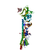
| |||||||||
|---|---|---|---|---|---|---|---|---|---|---|
| 1 | 
| |||||||||
| Unit cell |
| |||||||||
| Components on special symmetry positions |
|
-Components
-Protein , 1 types, 1 molecules A
| #1: Protein | Mass: 57131.680 Da / Num. of mol.: 1 / Fragment: Hemagglutinin Source method: isolated from a genetically manipulated source Source: (gene. exp.) Influenza A virus (strain A/Hong Kong/1/1968 H3N2)Strain: A/Hong Kong/1/1968 H3N2 / Gene: HA / Cell line (production host): HEK293 / Production host:  Homo sapiens (human) / References: UniProt: Q91MA7 Homo sapiens (human) / References: UniProt: Q91MA7 |
|---|
-Sugars , 4 types, 5 molecules 
| #2: Polysaccharide | beta-D-mannopyranose-(1-4)-2-acetamido-2-deoxy-beta-D-glucopyranose-(1-4)-2-acetamido-2-deoxy-beta- ...beta-D-mannopyranose-(1-4)-2-acetamido-2-deoxy-beta-D-glucopyranose-(1-4)-2-acetamido-2-deoxy-beta-D-glucopyranose Source method: isolated from a genetically manipulated source | ||
|---|---|---|---|
| #3: Polysaccharide | alpha-D-mannopyranose-(1-3)-[alpha-D-mannopyranose-(1-6)]beta-D-mannopyranose-(1-4)-2-acetamido-2- ...alpha-D-mannopyranose-(1-3)-[alpha-D-mannopyranose-(1-6)]beta-D-mannopyranose-(1-4)-2-acetamido-2-deoxy-beta-D-glucopyranose-(1-4)-2-acetamido-2-deoxy-beta-D-glucopyranose Source method: isolated from a genetically manipulated source | ||
| #4: Polysaccharide | Source method: isolated from a genetically manipulated source #5: Sugar | ChemComp-NAG / |  Type: D-saccharide, beta linking / Mass: 221.208 Da / Num. of mol.: 1 / Source method: obtained synthetically / Formula: C8H15NO6 Type: D-saccharide, beta linking / Mass: 221.208 Da / Num. of mol.: 1 / Source method: obtained synthetically / Formula: C8H15NO6 |
-Non-polymers , 2 types, 135 molecules 

| #6: Chemical | ChemComp-NO3 /  Mass: 62.005 Da / Num. of mol.: 1 / Source method: obtained synthetically / Formula: NO3 Mass: 62.005 Da / Num. of mol.: 1 / Source method: obtained synthetically / Formula: NO3 |
|---|---|
| #7: Water | ChemComp-HOH / Mass: 18.015 Da / Num. of mol.: 134 / Source method: isolated from a natural source / Formula: H2O |
-Details
| Has ligand of interest | N |
|---|---|
| Has protein modification | Y |
-Experimental details
-Experiment
| Experiment | Method: X-RAY DIFFRACTION / Number of used crystals: 2 |
|---|
- Sample preparation
| Crystal |
| |||||||||
|---|---|---|---|---|---|---|---|---|---|---|
| Crystal grow | Temperature: 293 K / Method: vapor diffusion / Details: 20.00 %w/v PEG 3350, 0.2 M LiNO3 |
-Data collection
| Diffraction | Mean temperature: 100 K / Serial crystal experiment: N |
|---|---|
| Diffraction source | Source: SYNCHROTRON / Site: SLS  / Beamline: X10SA / Wavelength: 0.999924 Å / Beamline: X10SA / Wavelength: 0.999924 Å |
| Detector | Type: DECTRIS EIGER X 16M / Detector: PIXEL / Date: Jan 28, 2021 |
| Radiation | Protocol: SINGLE WAVELENGTH / Monochromatic (M) / Laue (L): M / Scattering type: x-ray |
| Radiation wavelength | Wavelength: 0.999924 Å / Relative weight: 1 |
| Reflection | Resolution: 2.192→108.96 Å / Num. obs: 31315 / % possible obs: 100 % / Redundancy: 25.6 % / Rmerge(I) obs: 0.085 / Rsym value: 0.085 / Net I/σ(I): 24.9 |
| Reflection shell | Resolution: 2.192→108.96 Å / Redundancy: 26.2 % / Rmerge(I) obs: 2.977 / Mean I/σ(I) obs: 1.3 / Num. unique obs: 31315 / Rsym value: 2.977 / % possible all: 100 |
- Processing
| Software |
| |||||||||||||||||||||||||||||||||||||||||||||||||||||||||||||||||||||||||||
|---|---|---|---|---|---|---|---|---|---|---|---|---|---|---|---|---|---|---|---|---|---|---|---|---|---|---|---|---|---|---|---|---|---|---|---|---|---|---|---|---|---|---|---|---|---|---|---|---|---|---|---|---|---|---|---|---|---|---|---|---|---|---|---|---|---|---|---|---|---|---|---|---|---|---|---|---|
| Refinement | Method to determine structure: MOLECULAR REPLACEMENT Starting model: none Resolution: 2.19→108.96 Å / Cor.coef. Fo:Fc: 0.956 / Cor.coef. Fo:Fc free: 0.944 / SU B: 13.987 / SU ML: 0.176 / Cross valid method: THROUGHOUT / σ(F): 0 / ESU R: 0.246 / ESU R Free: 0.208 / Stereochemistry target values: MAXIMUM LIKELIHOOD Details: U VALUES : WITH TLS ADDED HYDROGENS HAVE BEEN ADDED IN THE RIDING POSITIONS
| |||||||||||||||||||||||||||||||||||||||||||||||||||||||||||||||||||||||||||
| Solvent computation | Ion probe radii: 0.8 Å / Shrinkage radii: 0.8 Å / VDW probe radii: 1.2 Å / Solvent model: MASK | |||||||||||||||||||||||||||||||||||||||||||||||||||||||||||||||||||||||||||
| Displacement parameters | Biso max: 158.29 Å2 / Biso mean: 66.571 Å2 / Biso min: 39.58 Å2
| |||||||||||||||||||||||||||||||||||||||||||||||||||||||||||||||||||||||||||
| Refinement step | Cycle: final / Resolution: 2.19→108.96 Å
| |||||||||||||||||||||||||||||||||||||||||||||||||||||||||||||||||||||||||||
| Refine LS restraints |
| |||||||||||||||||||||||||||||||||||||||||||||||||||||||||||||||||||||||||||
| LS refinement shell | Resolution: 2.192→2.249 Å / Rfactor Rfree error: 0 / Total num. of bins used: 20
| |||||||||||||||||||||||||||||||||||||||||||||||||||||||||||||||||||||||||||
| Refinement TLS params. | Method: refined / Refine-ID: X-RAY DIFFRACTION
| |||||||||||||||||||||||||||||||||||||||||||||||||||||||||||||||||||||||||||
| Refinement TLS group |
|
