 Movie
Movie Controller

[English] 日本語
 Yorodumi
- PDB-7q9x: Crystal structure of Chromobacterium violaceum aminotransferase i... -
+ Open data

- Basic information
| Entry | Database: PDB / ID: 7q9x | |||||||||
|---|---|---|---|---|---|---|---|---|---|---|
| Title | Crystal structure of Chromobacterium violaceum aminotransferase in complex with PLP-pyruvate adduct | |||||||||
 Components Components | Probable aminotransferase | |||||||||
 Keywords Keywords | TRANSFERASE / Transaminase / inhibition / pyridoxal phosphate | |||||||||
| Function / homology |  Function and homology information Function and homology informationadenosylmethionine-8-amino-7-oxononanoate transaminase / S-adenosyl-L-methionine:8-amino-7-oxononanoate transaminase activity / pyridoxal phosphate binding / identical protein binding / cytosol Similarity search - Function | |||||||||
| Biological species |  Chromobacterium violaceum (bacteria) Chromobacterium violaceum (bacteria) | |||||||||
| Method |  X-RAY DIFFRACTION / SYNCHROTRON / MOLECULAR REPLACEMENT / Resolution: 1.6 Å X-RAY DIFFRACTION / SYNCHROTRON / MOLECULAR REPLACEMENT / Resolution: 1.6 Å | |||||||||
 Authors Authors | Isupov, M.N. / Mitchell, D. / Sayer, C. / Littlechild, J.A. | |||||||||
| Funding support |  United Kingdom, 2items United Kingdom, 2items
| |||||||||
 Citation Citation | Journal: To Be Published Title: Aminotransferase from Chromobacterium violaceum in complex with PLP-pyruvate adduct. Authors: Isupov, M.N. / Mitchell, D. / Sayer, C. / Littlechild, J.A. | |||||||||
| History |
|
- Structure visualization
| Structure viewer | Molecule: MolmilJmol/JSmol |
|---|
- Downloads & links
-Download
| PDBx/mmCIF format | 7q9x.cif.gz | 440 KB | Display | PDBx/mmCIF format |
|---|---|---|---|---|
| PDB format | pdb7q9x.ent.gz | Display | PDB format | |
| PDBx/mmJSON format | 7q9x.json.gz | Tree view | PDBx/mmJSON format | |
| Others |  Other downloads Other downloads |
-Validation report
| Arichive directory | https://data.pdbj.org/pub/pdb/validation_reports/q9/7q9xftp://data.pdbj.org/pub/pdb/validation_reports/q9/7q9x | HTTPS FTP |
|---|
-Related structure data
| Related structure data |  7q9zC  4ba4S S: Starting model for refinement C: citing same article ( |
|---|---|
| Similar structure data |


-Links
 PDBj
PDBj- Assembly
| Deposited unit | 
| ||||||||||||||||||||||||||||||||||||||||||||||||||||||||||||||||||||||||||||||||||||||||||||||||||||||||||||||||||||||
|---|---|---|---|---|---|---|---|---|---|---|---|---|---|---|---|---|---|---|---|---|---|---|---|---|---|---|---|---|---|---|---|---|---|---|---|---|---|---|---|---|---|---|---|---|---|---|---|---|---|---|---|---|---|---|---|---|---|---|---|---|---|---|---|---|---|---|---|---|---|---|---|---|---|---|---|---|---|---|---|---|---|---|---|---|---|---|---|---|---|---|---|---|---|---|---|---|---|---|---|---|---|---|---|---|---|---|---|---|---|---|---|---|---|---|---|---|---|---|---|
| 1 | 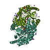
| ||||||||||||||||||||||||||||||||||||||||||||||||||||||||||||||||||||||||||||||||||||||||||||||||||||||||||||||||||||||
| 2 | 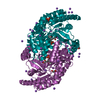
| ||||||||||||||||||||||||||||||||||||||||||||||||||||||||||||||||||||||||||||||||||||||||||||||||||||||||||||||||||||||
| Unit cell |
| ||||||||||||||||||||||||||||||||||||||||||||||||||||||||||||||||||||||||||||||||||||||||||||||||||||||||||||||||||||||
| Noncrystallographic symmetry (NCS) | NCS domain:
NCS domain segments: Beg auth comp-ID: ARG / Beg label comp-ID: ARG / End auth comp-ID: ALA / End label comp-ID: ALA / Auth seq-ID: 5 - 459 / Label seq-ID: 39 - 493
NCS ensembles :
|
-Components
-Protein , 1 types, 4 molecules AAABBBCCCDDD
| #1: Protein | Mass: 54832.230 Da / Num. of mol.: 4 Source method: isolated from a genetically manipulated source Source: (gene. exp.) Chromobacterium violaceum (strain ATCC 12472 / DSM 30191 / JCM 1249 / NBRC 12614 / NCIMB 9131 / NCTC 9757) (bacteria)Strain: ATCC 12472 / DSM 30191 / JCM 1249 / NBRC 12614 / NCIMB 9131 / NCTC 9757 Gene: CV_2025 / Production host: References: UniProt: Q7NWG4, adenosylmethionine-8-amino-7-oxononanoate transaminase |
|---|
-Non-polymers , 7 types, 2121 molecules 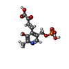


| #2: Chemical | ChemComp-AN7 / (  Mass: 317.189 Da / Num. of mol.: 4 / Source method: obtained synthetically / Formula: C11H12NO8P / Feature type: SUBJECT OF INVESTIGATION Mass: 317.189 Da / Num. of mol.: 4 / Source method: obtained synthetically / Formula: C11H12NO8P / Feature type: SUBJECT OF INVESTIGATION#3: Chemical | ChemComp-EDO /  Mass: 62.068 Da / Num. of mol.: 5 / Source method: obtained synthetically / Formula: C2H6O2 Mass: 62.068 Da / Num. of mol.: 5 / Source method: obtained synthetically / Formula: C2H6O2#4: Chemical | ChemComp-NA /  Mass: 22.990 Da / Num. of mol.: 77 / Source method: obtained synthetically / Formula: Na Mass: 22.990 Da / Num. of mol.: 77 / Source method: obtained synthetically / Formula: Na#5: Chemical | ChemComp-PLP / |  Mass: 247.142 Da / Num. of mol.: 1 / Source method: obtained synthetically / Formula: C8H10NO6P / Feature type: SUBJECT OF INVESTIGATION Mass: 247.142 Da / Num. of mol.: 1 / Source method: obtained synthetically / Formula: C8H10NO6P / Feature type: SUBJECT OF INVESTIGATION#6: Chemical |  Mass: 94.971 Da / Num. of mol.: 2 / Source method: obtained synthetically / Formula: PO4 Mass: 94.971 Da / Num. of mol.: 2 / Source method: obtained synthetically / Formula: PO4#7: Chemical | ChemComp-PEG / |  Mass: 106.120 Da / Num. of mol.: 1 / Source method: isolated from a natural source / Formula: C4H10O3 Mass: 106.120 Da / Num. of mol.: 1 / Source method: isolated from a natural source / Formula: C4H10O3#8: Water | ChemComp-HOH / | Mass: 18.015 Da / Num. of mol.: 2031 / Source method: isolated from a natural source / Formula: H2O |
|---|
-Details
| Has ligand of interest | Y |
|---|
-Experimental details
-Experiment
| Experiment | Method: X-RAY DIFFRACTION / Number of used crystals: 1 |
|---|
- Sample preparation
| Crystal | Density Matthews: 1.99 Å3/Da / Density % sol: 38.12 % |
|---|---|
| Crystal grow | Temperature: 290 K / Method: microbatch Details: 12.5% PEG 8000 and 50 mM HEPES pH 7.5 10 mM PLP, 5 mM pyruvate |
-Data collection
| Diffraction | Mean temperature: 100 K / Serial crystal experiment: N |
|---|---|
| Diffraction source | Source: SYNCHROTRON / Site: Diamond / Beamline: I03 / Wavelength: 0.97 Å |
| Detector | Type: ADSC QUANTUM 315 / Detector: CCD / Date: Jun 23, 2008 |
| Radiation | Protocol: SINGLE WAVELENGTH / Monochromatic (M) / Laue (L): M / Scattering type: x-ray |
| Radiation wavelength | Wavelength: 0.97 Å / Relative weight: 1 |
| Reflection | Resolution: 1.6→39.43 Å / Num. obs: 156999 / % possible obs: 76.1 % / Redundancy: 4.7 % / CC1/2: 0.999 / Net I/σ(I): 14.9 |
| Reflection shell | Resolution: 1.6→1.64 Å / Redundancy: 4.3 % / Mean I/σ(I) obs: 2.5 / Num. unique obs: 3529 / CC1/2: 0.861 / % possible all: 23 |
- Processing
| Software |
| |||||||||||||||||||||||||||||||||||||||||||||||||||||||||||||||||||||||||||||||||||||||||||||||||||||||||||||||||||||||||||||||||||||||||||||||||||
|---|---|---|---|---|---|---|---|---|---|---|---|---|---|---|---|---|---|---|---|---|---|---|---|---|---|---|---|---|---|---|---|---|---|---|---|---|---|---|---|---|---|---|---|---|---|---|---|---|---|---|---|---|---|---|---|---|---|---|---|---|---|---|---|---|---|---|---|---|---|---|---|---|---|---|---|---|---|---|---|---|---|---|---|---|---|---|---|---|---|---|---|---|---|---|---|---|---|---|---|---|---|---|---|---|---|---|---|---|---|---|---|---|---|---|---|---|---|---|---|---|---|---|---|---|---|---|---|---|---|---|---|---|---|---|---|---|---|---|---|---|---|---|---|---|---|---|---|---|
| Refinement | Method to determine structure: MOLECULAR REPLACEMENT Starting model: 4ba4 Resolution: 1.6→39.43 Å / Cor.coef. Fo:Fc: 0.955 / Cor.coef. Fo:Fc free: 0.94 / Cross valid method: FREE R-VALUE / ESU R: 0.187 / ESU R Free: 0.158 / Details: Hydrogens have not been used
| |||||||||||||||||||||||||||||||||||||||||||||||||||||||||||||||||||||||||||||||||||||||||||||||||||||||||||||||||||||||||||||||||||||||||||||||||||
| Solvent computation | Ion probe radii: 0.8 Å / Shrinkage radii: 0.8 Å / VDW probe radii: 1.2 Å / Solvent model: BABINET MODEL PLUS MASK | |||||||||||||||||||||||||||||||||||||||||||||||||||||||||||||||||||||||||||||||||||||||||||||||||||||||||||||||||||||||||||||||||||||||||||||||||||
| Displacement parameters | Biso mean: 17.71 Å2
| |||||||||||||||||||||||||||||||||||||||||||||||||||||||||||||||||||||||||||||||||||||||||||||||||||||||||||||||||||||||||||||||||||||||||||||||||||
| Refinement step | Cycle: LAST / Resolution: 1.6→39.43 Å
| |||||||||||||||||||||||||||||||||||||||||||||||||||||||||||||||||||||||||||||||||||||||||||||||||||||||||||||||||||||||||||||||||||||||||||||||||||
| Refine LS restraints |
| |||||||||||||||||||||||||||||||||||||||||||||||||||||||||||||||||||||||||||||||||||||||||||||||||||||||||||||||||||||||||||||||||||||||||||||||||||
| Refine LS restraints NCS |
| |||||||||||||||||||||||||||||||||||||||||||||||||||||||||||||||||||||||||||||||||||||||||||||||||||||||||||||||||||||||||||||||||||||||||||||||||||
| LS refinement shell |
|
