 Movie
Movie Controller

[English] 日本語
 Yorodumi
- PDB-7q8e: Crystal Structure of the MurT-GatD Enzyme Complex from Staphyloco... -
+ Open data

- Basic information
| Entry | Database: PDB / ID: 7q8e | |||||||||||||||
|---|---|---|---|---|---|---|---|---|---|---|---|---|---|---|---|---|
| Title | Crystal Structure of the MurT-GatD Enzyme Complex from Staphylococcus aureus COL strain | |||||||||||||||
 Components Components |
| |||||||||||||||
 Keywords Keywords | LIGASE / Essential Glutamine Amidotransferase / Glutaminase/Ligase / Bacterial Peptidoglycan Amidation. | |||||||||||||||
| Function / homology |  Function and homology information Function and homology informationlipid II isoglutaminyl synthase (glutamine-hydrolysing) / carbon-nitrogen ligase activity on lipid II / acid-amino acid ligase activity / glutaminase / cobalamin biosynthetic process / glutaminase activity / peptidoglycan biosynthetic process / cell wall organization / regulation of cell shape / zinc ion binding / ATP binding Similarity search - Function | |||||||||||||||
| Biological species |  Staphylococcus aureus subsp. aureus COL (bacteria) Staphylococcus aureus subsp. aureus COL (bacteria) | |||||||||||||||
| Method |  X-RAY DIFFRACTION / SYNCHROTRON / MOLECULAR REPLACEMENT / Resolution: 2.90001720351 Å X-RAY DIFFRACTION / SYNCHROTRON / MOLECULAR REPLACEMENT / Resolution: 2.90001720351 Å | |||||||||||||||
 Authors Authors | Leisico, F. / Romao, M.J. / Santos-Silva, T. | |||||||||||||||
| Funding support |  Portugal, 4items Portugal, 4items
| |||||||||||||||
 Citation Citation | Journal: To Be Published Title: Crystal Structure of the MurT-GatD Enzyme Complex from Staphylococcus aureus COL strain Authors: Leisico, F. / Romao, M.J. / Santos-Silva, T. | |||||||||||||||
| History |
|
- Structure visualization
| Structure viewer | Molecule: MolmilJmol/JSmol |
|---|
- Downloads & links
-Download
| PDBx/mmCIF format | 7q8e.cif.gz | 611.1 KB | Display | PDBx/mmCIF format |
|---|---|---|---|---|
| PDB format | pdb7q8e.ent.gz | 423.9 KB | Display | PDB format |
| PDBx/mmJSON format | 7q8e.json.gz | Tree view | PDBx/mmJSON format | |
| Others |  Other downloads Other downloads |
-Validation report
| Arichive directory | https://data.pdbj.org/pub/pdb/validation_reports/q8/7q8eftp://data.pdbj.org/pub/pdb/validation_reports/q8/7q8e | HTTPS FTP |
|---|
-Related structure data
| Related structure data |  6gs2S S: Starting model for refinement |
|---|---|
| Similar structure data | |
| Other databases |
|
-Links
 PDBj
PDBj

- Assembly
| Deposited unit | 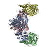
| |||||||||||||||||||||||||||||||||||||||||||||||||||||||||||||||||||||||||
|---|---|---|---|---|---|---|---|---|---|---|---|---|---|---|---|---|---|---|---|---|---|---|---|---|---|---|---|---|---|---|---|---|---|---|---|---|---|---|---|---|---|---|---|---|---|---|---|---|---|---|---|---|---|---|---|---|---|---|---|---|---|---|---|---|---|---|---|---|---|---|---|---|---|---|
| 1 | 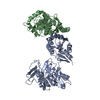
| |||||||||||||||||||||||||||||||||||||||||||||||||||||||||||||||||||||||||
| 2 | 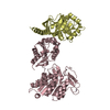
| |||||||||||||||||||||||||||||||||||||||||||||||||||||||||||||||||||||||||
| Unit cell |
| |||||||||||||||||||||||||||||||||||||||||||||||||||||||||||||||||||||||||
| Noncrystallographic symmetry (NCS) | NCS domain:
NCS domain segments:
NCS ensembles :
|
-Components
| #1: Protein | Mass: 49264.910 Da / Num. of mol.: 2 Source method: isolated from a genetically manipulated source Source: (gene. exp.) Staphylococcus aureus subsp. aureus COL (bacteria)Gene: murT, SACOL1951 / Production host: References: UniProt: A0A0H2WZQ7, lipid II isoglutaminyl synthase (glutamine-hydrolysing) #2: Protein | Mass: 28539.184 Da / Num. of mol.: 2 Source method: isolated from a genetically manipulated source Source: (gene. exp.) Staphylococcus aureus subsp. aureus COL (bacteria)Gene: gatD, SACOL1950 / Production host: References: UniProt: A0A0H2WZ38, lipid II isoglutaminyl synthase (glutamine-hydrolysing), glutaminase #3: Chemical |   Mass: 65.409 Da / Num. of mol.: 2 / Source method: obtained synthetically / Formula: Zn / Feature type: SUBJECT OF INVESTIGATION Mass: 65.409 Da / Num. of mol.: 2 / Source method: obtained synthetically / Formula: Zn / Feature type: SUBJECT OF INVESTIGATION#4: Chemical | ChemComp-CL / |   Mass: 35.453 Da / Num. of mol.: 1 / Source method: obtained synthetically / Formula: Cl Mass: 35.453 Da / Num. of mol.: 1 / Source method: obtained synthetically / Formula: Cl#5: Water | ChemComp-HOH / |  Mass: 18.015 Da / Num. of mol.: 75 / Source method: isolated from a natural source / Formula: H2O Mass: 18.015 Da / Num. of mol.: 75 / Source method: isolated from a natural source / Formula: H2OHas ligand of interest | Y | |
|---|
-Experimental details
-Experiment
| Experiment | Method: X-RAY DIFFRACTION / Number of used crystals: 1 |
|---|
- Sample preparation
| Crystal | Density Matthews: 2.09 Å3/Da / Density % sol: 42.76 % |
|---|---|
| Crystal grow | Temperature: 293 K / Method: vapor diffusion, sitting drop Details: 30 % (w/v) PEG 4,000, 100 mM Tris-HCl pH 8.5 and 200 mM MgCl2; |
-Data collection
| Diffraction | Mean temperature: 100 K / Serial crystal experiment: N |
|---|---|
| Diffraction source | Source: SYNCHROTRON / Site: ESRF  / Beamline: ID23-2 / Wavelength: 0.8731 Å / Beamline: ID23-2 / Wavelength: 0.8731 Å |
| Detector | Type: DECTRIS PILATUS 2M / Detector: PIXEL / Date: Jul 13, 2018 |
| Radiation | Protocol: SINGLE WAVELENGTH / Monochromatic (M) / Laue (L): M / Scattering type: x-ray |
| Radiation wavelength | Wavelength: 0.8731 Å / Relative weight: 1 |
| Reflection | Resolution: 2.9→49.31 Å / Num. obs: 30388 / % possible obs: 99.99 % / Redundancy: 6.9 % / Biso Wilson estimate: 62.9479579287 Å2 / CC1/2: 0.99 / Rpim(I) all: 0.113 / Net I/σ(I): 6.7 |
| Reflection shell | Resolution: 2.9→3.06 Å / Redundancy: 7.4 % / Mean I/σ(I) obs: 17 / Num. unique obs: 4366 / CC1/2: 0.58 / Rpim(I) all: 0.696 / % possible all: 100 |
- Processing
| Software |
| |||||||||||||||||||||||||||||||||||||||||||||||||||||||||||||||||||||||||||||||||||||||||||||||||||||||||||||||||||||||||||||
|---|---|---|---|---|---|---|---|---|---|---|---|---|---|---|---|---|---|---|---|---|---|---|---|---|---|---|---|---|---|---|---|---|---|---|---|---|---|---|---|---|---|---|---|---|---|---|---|---|---|---|---|---|---|---|---|---|---|---|---|---|---|---|---|---|---|---|---|---|---|---|---|---|---|---|---|---|---|---|---|---|---|---|---|---|---|---|---|---|---|---|---|---|---|---|---|---|---|---|---|---|---|---|---|---|---|---|---|---|---|---|---|---|---|---|---|---|---|---|---|---|---|---|---|---|---|---|
| Refinement | Method to determine structure: MOLECULAR REPLACEMENT Starting model: 6GS2 Resolution: 2.90001720351→49.3089437971 Å / SU ML: 0.442344360262 / Cross valid method: FREE R-VALUE / σ(F): 1.33648315019 / Phase error: 30.4477447237 Stereochemistry target values: GeoStd + Monomer Library + CDL v1.2
| |||||||||||||||||||||||||||||||||||||||||||||||||||||||||||||||||||||||||||||||||||||||||||||||||||||||||||||||||||||||||||||
| Solvent computation | Shrinkage radii: 0.9 Å / VDW probe radii: 1.11 Å / Solvent model: FLAT BULK SOLVENT MODEL | |||||||||||||||||||||||||||||||||||||||||||||||||||||||||||||||||||||||||||||||||||||||||||||||||||||||||||||||||||||||||||||
| Displacement parameters | Biso mean: 73.5780373074 Å2 | |||||||||||||||||||||||||||||||||||||||||||||||||||||||||||||||||||||||||||||||||||||||||||||||||||||||||||||||||||||||||||||
| Refinement step | Cycle: LAST / Resolution: 2.90001720351→49.3089437971 Å
| |||||||||||||||||||||||||||||||||||||||||||||||||||||||||||||||||||||||||||||||||||||||||||||||||||||||||||||||||||||||||||||
| Refine LS restraints |
| |||||||||||||||||||||||||||||||||||||||||||||||||||||||||||||||||||||||||||||||||||||||||||||||||||||||||||||||||||||||||||||
| LS refinement shell |
| |||||||||||||||||||||||||||||||||||||||||||||||||||||||||||||||||||||||||||||||||||||||||||||||||||||||||||||||||||||||||||||
| Refinement TLS params. | Method: refined / Refine-ID: X-RAY DIFFRACTION
| |||||||||||||||||||||||||||||||||||||||||||||||||||||||||||||||||||||||||||||||||||||||||||||||||||||||||||||||||||||||||||||
| Refinement TLS group |
|
